
Stem cell introduction device

An introduction device and stem cell technology, which can be used in drug devices and other medical devices, etc., can solve problems such as the delivery of stem cells to lesions, and achieve the effects of being convenient for clinical promotion, economical and practical, and improving reproductive function.

Active Publication Date: 2016-04-13
PEKING UNIV SHENZHEN HOSPITAL
View PDF8 Cites 0 Cited by
  • Summary
  • Abstract
  • Description
  • Claims
  • Application Information

AI Technical Summary

Problems solved by technology

In the field of obstetrics and gynecology, for clinical difficulties such as thin endometrium, endometrial lesions or injuries, local introduction of stem cells is a new treatment method to improve the local environment of the endometrium and promote endometrial repair. Clinically, it is necessary to solve the problem of introducing stem cells into the Intimal basal approach, however, how to deliver stem cells to the lesion site is an unresolved question

Method used


Image

  • Stem cell introduction device
  • Stem cell introduction device

Examples


Embodiment 1

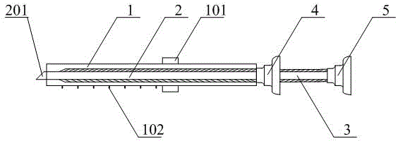
[0023] See figure 1 , the stem cell introducing device comprises a hollow outer tube 1 , a hollow needle core 2 and a hollow catheter 3 . The outer tube 1 is a straight round tube with a blunt and smooth top made of medical non-toxic hard plastic material. The wall thickness of the tube is 1.0 mm, the outer diameter is 5.5 mm, and the length is 15 cm; 1. A cursor 101 that moves up and down on the outer wall. On the other side of the outer wall of the outer sleeve 1, a digital scale mark 102 protruding from the surface of the outer wall of the outer sleeve 1 is provided every 0.5 cm downward with the top of the outer sleeve as the zero point, until 9 cm. , when the vernier 101 moves to the digital scale mark 102 , then rotate clockwise or counterclockwise by 90°~180° to rotate the vernier 101 to fix it on the digital scale mark 102 . The needle core 2 is a straight round tube made of metal material, with an outer diameter of 3 mm, an inner diameter of 1 mm, and a length of 15....

Embodiment 2

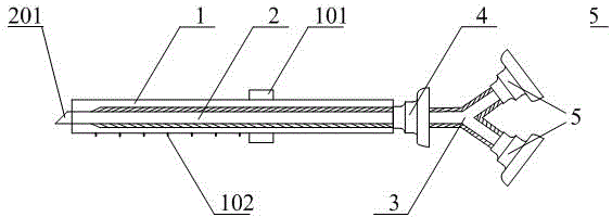
[0025] See figure 2 , figure 2 It is a sectional view of a stem cell introduction device provided by another embodiment of the present invention. The outer sleeve, needle core, first syringe interface and second syringe interface of the stem cell introduction device provided in this embodiment are connected with figure 1 The outer cannula, needle core, first syringe interface and second syringe interface of the stem cell introduction device provided by the illustrated embodiment are the same, and are the same as figure 1The difference between the stem cell introduction device provided by the illustrated embodiment is that the catheter 3 is set as a Y-shaped three-way pipe, the main pipe at the proximal end of the Y-shaped three-way pipe communicates with the other end of the first syringe interface 4, and the distal end of the Y-shaped three-way pipe The two side pipes of the two side pipes are respectively connected to a second syringe interface 5, and the second syringe i...


PUM


Abstract

The invention discloses a stem cell introduction device. The stem cell introduction device comprises a hollow outer cannula, a hollow needle core and a hollow catheter. A vernier is arranged along one side on the outer wall of the outer cannula. The other side of the outer wall of the hollow outer cannula is provided with digit scale marks protruding from the outer wall surface. When moving to the digit scale mark, the vernier is rotated to be secured between digit scale marks or above them. The head of the needle core is provided with a piercing part. The tail of the needle core is connected with one end of a first injector joint. The piercing part is exposed by two millimeters when the needle core is completely inserted into the hollow part of the outer cannula. The near end of the cannula is connected to the other end of the first injector joint. The distal end of the cannula is connected to a second injector joint. The stem cell introduction device has following beneficial effects: the stem cell introduction device can accurately introduce stem cells into the stratum basale of the endometrium such that the damaged endometrium of a patient can be repaired; the reproduction function is improved so that diseases related to endometria can be treated; and the stem cell introduction device is easy and convenient to operate, cost-effective and practical and is convenient for clinic use.

Description

technical field [0001] The invention relates to a stem cell introduction device, in particular to an introduction device for introducing stem cells or medicine into the basal layer of the endometrium. Background technique [0002] The endometrium is the organ with the most active growth in the human body. It is divided into a functional layer and a basal layer. The former proliferates and changes with the changes of estrogen and progesterone in the menstrual cycle; the latter repairs wounds and hyperplasia after the endometrial shedding. Form a new functional layer. The source of this repair process of endometrial wounds during menstruation is the stem cells located in the basal layer of the endometrium. In the past, it was believed that the damage and destruction of the basal layer of the endometrium were irreversible lesions, which could cause amenorrhea and infertility in patients. In animal experiments, stem cells were injected into the junction of the myometrium and e...

Claims


Application Information

Patent Timeline
Patent Type & Authority Applications(China)
IPC IPC(8): A61M31/00
Inventor 吴瑞芳汤惠茹王国平杜辉
Owner PEKING UNIV SHENZHEN HOSPITAL
Learn More