Method and device for obtaining biophysical characteristics of ultra-diffraction limit cell membrane micro-structure

A super-diffraction limit and biophysical technology, applied in the field of super-diffraction limit cell membrane microstructure biophysical characteristics acquisition and device, can solve the problem that the atomic force probe has no specific recognition ability, does not have the ability to obtain nano-scale ultrastructure functional information, etc. question

Active Publication Date: 2016-05-11
FUJIAN NORMAL UNIV
View PDF7 Cites 11 Cited by
  • Summary
  • Abstract
  • Description
  • Claims
  • Application Information

AI Technical Summary

Problems solved by technology

However, atomic force probes have no specific recognition ability, while SIM does not have the ability to obtain functional information related to nanoscale ultrastructure.

Method used


Image

  • Method and device for obtaining biophysical characteristics of ultra-diffraction limit cell membrane micro-structure
  • Method and device for obtaining biophysical characteristics of ultra-diffraction limit cell membrane micro-structure
  • Method and device for obtaining biophysical characteristics of ultra-diffraction limit cell membrane micro-structure

Examples


Embodiment Construction

[0047] In order to make the above-mentioned features and advantages of the present invention more comprehensible, the following specific embodiments are described in detail with reference to the accompanying drawings.

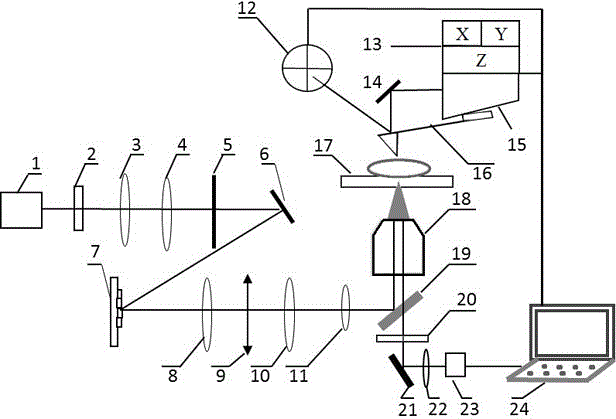
[0048] Such as figure 1 As shown, a device for acquiring biophysical properties of super-diffraction-limited cell membrane microstructures includes a laser 1 and a semiconductor laser 15. The laser output optical path of the laser 1 is sequentially provided with a polarizer 2, a first converging lens group, an aperture 5, The first reflecting mirror 6, the spatial light modulator 7, the third converging lens 8, the light blocking plate 9, the second converging lens group and the dichroic mirror 19, the reflected light output optical path of the dichroic mirror 19 is provided with optical The objective lens 18 and the stage 17 of the microscope, the transmitted light output optical path of the dichroic mirror 19 is provided with a filter plate 20, a switchable m...


PUM


Abstract

The invention relates to a method and device for obtaining the biophysical characteristics of an ultra-diffraction limit cell membrane micro-structure. The device comprises a laser device and a semiconductor laser device. The laser output path of the laser device sequentially comprises: a polarizer, a first convergent lens set, an aperture, a first reflector, a spatial light modulator, a third convergent lens, a light barrier, a second convergent lest set, and a dichroscope; the reflected light output path of the dichroscope sequentially comprises the objective lens and the objective table of an optical microscope; the transmission light output path of the dichroscope sequentially comprises a filter, a switchable reflector, a sixth convergent lens, and a CCD detector; the laser output path of the semiconductor laser device sequentially comprises a second reflector, a probe, which is driven by a three dimensional controller, and a position sensitive detector; and the CCD detector, the three dimensional controller, and the position sensitive detector are all connected to a data collecting and controlling system. The provided method and device can obtain the information of biophysical characteristics of an ultra-diffraction limit cell membrane micro-structure.

Description

technical field [0001] The invention relates to the coupling of different modal imaging technologies of optical microscopic imaging and atomic force scanning imaging, in particular to a method and device for acquiring biophysical properties of super-diffraction-limited cell membrane microstructures. Background technique [0002] The observation of specific and related biological processes on the cell membrane at the micro-nano scale has become a hot spot in recent years. Ordinary fluorescence optical microscopy occupies an important position in live cell microscopy and detection research due to its non-destructive characteristics to samples. However, since fluorescence microscopy, including laser confocal scanning microscopy, is limited by the diffraction limit, the spatial resolution can only approach half a wavelength. In order to break through the diffraction limit (super-diffraction limit), that is, the spatial resolution is below 200nm, in recent years various super-res...

Claims


Application Information

Patent Timeline
Patent Type & Authority Applications(China)
IPC IPC(8): G01N21/01G01Q60/02
CPCG01N21/01G01N2021/0112G01Q60/02
Inventor 王瑜华杨洪钦邱彩敏蒋宁城尤明海陈建玲谢树森
Owner FUJIAN NORMAL UNIV
Why Eureka
Try Eureka