Method and system automatically detecting local lesion in radiographic image

A localized, radiological image technology, applied in the field of re-screening and automatic abnormal inspection, which can solve problems such as difficult to correct errors, achieve the effect of increasing accuracy and eliminating false positives

Inactive Publication Date: 2016-06-08
SHENZHEN SMART IMAGING HEALTHCARE
View PDF3 Cites 26 Cited by
  • Summary
  • Abstract
  • Description
  • Claims
  • Application Information

AI Technical Summary

Problems solved by technology

Those experienced lung radiologists can achieve a high degree of accuracy in diagnosis. However, in the training stage of doctors, although they have a high level of clin

Method used


Image

  • Method and system automatically detecting local lesion in radiographic image
  • Method and system automatically detecting local lesion in radiographic image
  • Method and system automatically detecting local lesion in radiographic image

Examples


Embodiment Construction

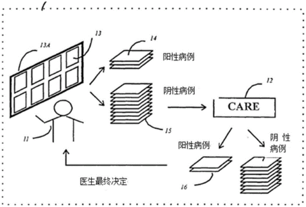
[0021] figure 1 Denotes a diagnostic step to improve the detection of suspicious focal lesions. The X-ray chest film 13 of the patient is first installed in the light box 13A of the radiation device. The radiologist carefully examines the possible localized pulmonary lesions in the image 13, and the radiologist confirms that there are suspicious localized pulmonary lesions. The image was defined as a positive image14 for further radiological diagnosis. Negative images 15 determined by the radiologist not to contain suspicious focal lesions are transmitted to the computer-aided re-screening (CARE) system 12 of the present invention for further diagnosis.

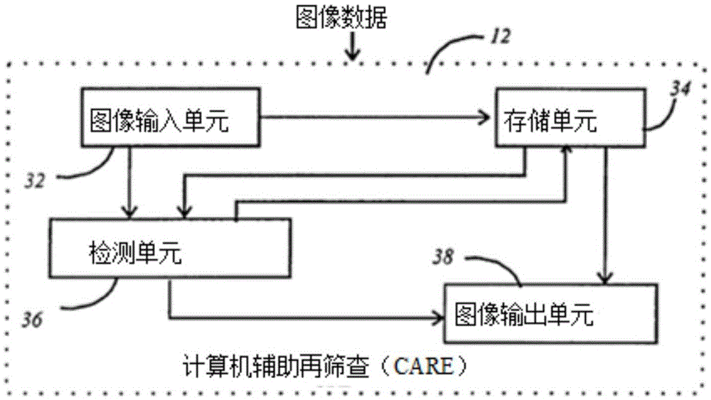
[0022] Computer Aided Rescreening (CARE) system 12 is computer based and involves a multi-stage process. There are two types of case confirmation in the CARE system12: positive cases16 and negative cases17. Positive case 16 was sent back to the radiologist for final decision based on chest x-rays.

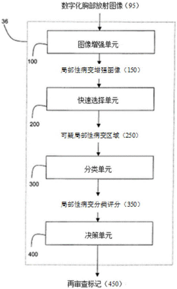
[0023] The method and ...


PUM


Abstract

An automatic detection method and system capable of improving radiographic image abnormal (lung cancer local lesion) detection process in the prior art is provided. The detection method and system employs multi-resolution technology to improve detection efficiency for local lesion with various sizes and further use virtual local lesion to conduct relevance and match, so local lesions of all or most sizes can be detected. The detection method and system employs spherical parameter to represent local lesions, so non-obvious local lesions can be accurately detected. A plurality of classifiers employed by the detection method and system comprise a counter-propagation neural network, data fusion, trimming neural network based on decision-making and convolution neural network structure, so classification grade of lung local lesion classification can be generated; and images with high dubiety can be further detected upon final decision-making.

Description

1. Technical field [0001] The present invention is used for a method and system for digital image processing, more specifically for a method and system for re-screening and abnormal automatic inspection, for example: multi-resolution processing of radioactive chest images with localized lung lesions, digital image processing and deep learning Neural Networks. 2. Background technology [0002] Lung cancer is the leading cancer type in both men and women worldwide, and early detection and treatment of potentially treatable stage localized lung cancer can significantly improve patient survival. Studies have shown that the probability of lung cancer being diagnosed by one radiologist is close to 68%, and the probability increases to 82% when a second reader is added. A long-term lung cancer screening program conducted at the Mayo Clinic found that 90% of small peripheral lung cancers could be detected by tracing chest X-rays of early lung cancers. [0003] Compared with common...

Claims


Application Information

Patent Timeline
IPC IPC(8): A61B6/00
CPCA61B6/5217
Inventor 刘远明权申文段淑婷周浩
Owner SHENZHEN SMART IMAGING HEALTHCARE
Why Eureka
Try Eureka