Separation and extraction method for cytomembrane microvesicles (MVs) and exosomes (EXs)

An extraction method and technology for microvesicles, which are applied in the field of medical invention and can solve problems such as research and development limitations

Inactive Publication Date: 2016-06-29
WUHAN DAFU BIOTECH CO LTD
View PDF3 Cites 13 Cited by
  • Summary
  • Abstract
  • Description
  • Claims
  • Application Information

AI Technical Summary

Problems solved by technology

However, traditional methods are difficult to sort and detect specific EVs, such as cEC-EVs and cEPC-EVs, in complex samples
Therefore, the development of research in this area is greatly limited

Method used


Image

  • Separation and extraction method for cytomembrane microvesicles (MVs) and exosomes (EXs)
  • Separation and extraction method for cytomembrane microvesicles (MVs) and exosomes (EXs)
  • Separation and extraction method for cytomembrane microvesicles (MVs) and exosomes (EXs)

Examples


Embodiment Construction

[0017] The following is a specific implementation case of this project. The reagents and materials used in the subordinate applications can be obtained through market purchase channels. The pH of PBS used in the present invention is 7.2.

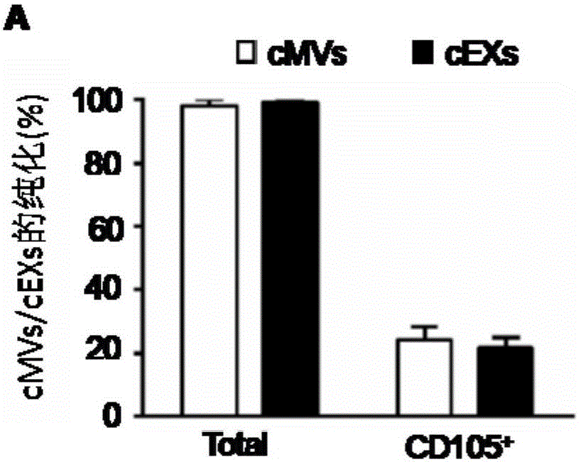
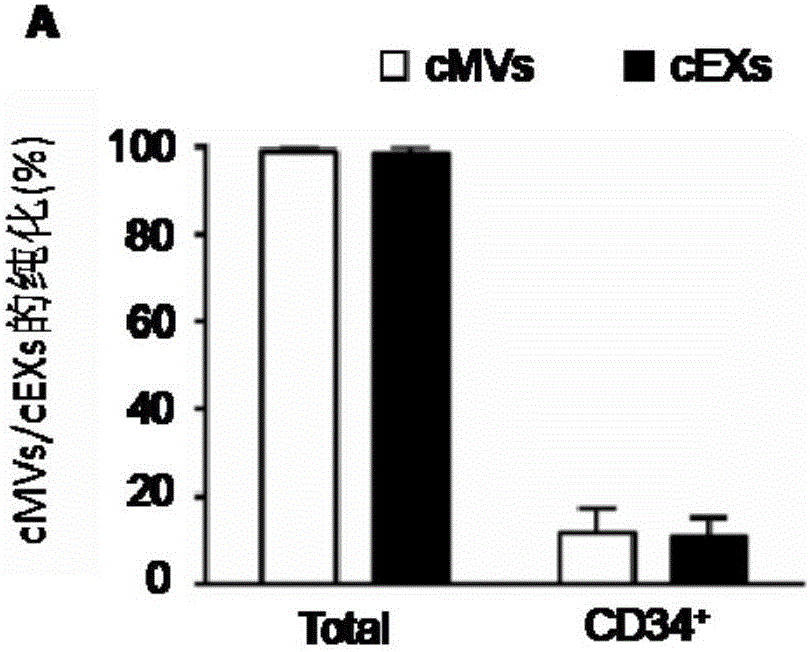
[0018] Such as Figure 4 As shown, extract 3ml of peripheral blood and put it in a 2ml sterile tube containing 3.3% sodium citrate; add 6ml of filtered PBS; mix well and perform centrifugation. Separation conditions: speed 400r / s, 35min, 4 ℃; take the uppermost liquid as plasma; take 1ml of plasma for centrifugation, separation conditions: speed 2000r / s, 20min, 4℃, used to remove platelets; take supernatant for centrifugation, separation conditions: speed 20000r / s, 120min, 4°C, the obtained precipitate was suspended in 700μl filtered PBS for NTA analysis of the total number of cMVs; the remaining supernatant was centrifuged, separation conditions: 169000r / s, 6h, 4°C, the obtained precipitate was used 700 μl of filtered PBS was suspended f...


PUM

PropertyMeasurementUnit
diameteraaaaaaaaaa
diameteraaaaaaaaaa

Abstract

The invention relates to a separation and extraction method for cytomembrane microvesicles (MVs) and exosomes (EXs). The method is characterized by comprising the following steps that a, 3-10 ml of a peripheral blood sample is drawn and placed in an anticoagulation tube to be stored, the sample is diluted with phosphate buffer normal saline (PBS) by 2-4 times, and then centrifugation is carried out to obtain supernate plasma; b, the supernate plasma is subjected to centrifugal separation again, and sediment is MVs; c, ultra-centrifugation is carried out on the supernate plasma in the step b, and sediment is EXs. According to the method, a previous method adopting mono-specificity antibodies is broken through, and the specificity and sensibility of the extracted cell MVs and EXs are higher than those in the prior art. A specificity and sensibility direction is provided for a further study of the MVs and the EXs as biomarkers of diseases.

Description

technical field [0001] The invention relates to a method for separating and extracting cell membrane microvesicles MVs and exosomes EXs, and belongs to the field of medical inventions. Background technique [0002] Extracellular vesicles (Extracellular vesicles, EVs) are a type of biologically active small vesicles secreted and released by cells. EVs can carry and transfer the lipids, functional proteins, messenger RNAs (mRNAs) and micro RNAs (miRNAs) of the mother cell into the target cells, and regulate the gene expression and function of the target cells. Due to the limitations of technical level and understanding, when researchers first proposed the concept of EVs, they only regarded EVs as "garbage bags" for cells, allowing cells to get rid of some useless protein garbage. Further research in the past ten years has found that the "cargo" carried by EVs has important biological significance, especially the miRNAs contained in it have been confirmed to be involved in the...

Claims


Application Information

Patent Timeline
Patent Type & Authority Applications(China)
IPC IPC(8): G01N1/34
CPCG01N1/34
Inventor 陈颜芳肖湘王景菊陈姬柳华
Owner WUHAN DAFU BIOTECH CO LTD
Why Eureka
Try Eureka