Eureka AIR delivers breakthrough ideas for toughest innovation challenges, trusted by R&D personnel around the world.

Density guided attenuation map generation in PET/MR systems

A dense, modular system technology used in medical imaging to solve problems such as incorrect segmentation

Inactive Publication Date: 2016-08-10
KONINKLJIJKE PHILIPS NV
View PDF2 Cites 9 Cited by
  • Summary
  • Abstract
  • Description
  • Claims
  • Application Information

AI Technical Summary

Problems solved by technology

Segmenting the lung as a whole for the purpose of generating an attenuation correction map may not be correct if there are lesions inside the lung

Method used


Image

  • Density guided attenuation map generation in PET/MR systems
  • Density guided attenuation map generation in PET/MR systems
  • Density guided attenuation map generation in PET/MR systems

Examples


Embodiment Construction

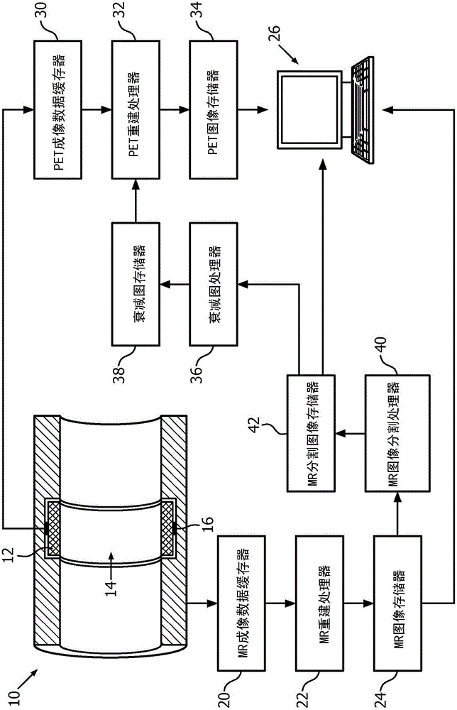
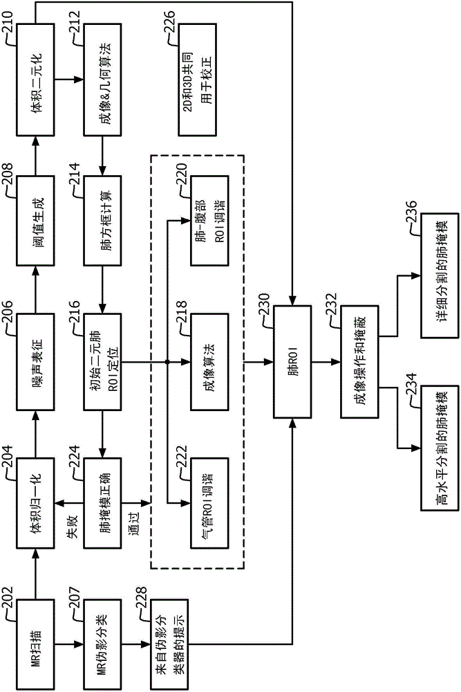
[0021] The present application provides accurate attenuation coefficients for internal tissue within the lung based on lung density comparison with segmented tissue. The present application provides a technique to classify each magnetic resonance (MR) scan based on noise characteristics. Noise characterization helps determine the feasibility of detailed and accurate lung segmentation. The present application is able to generate a lung region of interest (ROI) from the ROI as well as a detailed structural segmentation of the lung. The present application provides an iterative normalization and region definition method that accurately captures the entire lung and the soft tissue within the lung. The accuracy of the segmentation also comes from the classification of artifacts in the MR images. The present application proposes to correlate segmented lung interior tissue pixels with lung density to determine an attenuation coefficient based on the correlation. Pixels are 2D or 3...


PUM


Abstract

A lung segmentation processor (40) is configured to classify magnetic resonance (MR) images based on noise characteristics. The MR segmenatation processor generates a lung region of interest (ROI) and detailed structure segmentation of the lung from the ROI. The MR segmentation processor performs an iterative normalization and region definition approach that captures the entire lung and the soft tissues within the lung accurately. Accuracy of the segmentation relies on artifact classification coming inherently from MR images. The MR segmentation processor (40) correlates segmented lung internal tissue pixels with the lung density to determine the attenuation coefficients based on the correlation. Lung densities are computed by using MR data obtained from imaging sequences that minimize echo and acquisition times. The densities differentiate healthy tissues and lesions, which an attenuation map processor (36) uses to create localized attenuation maps for the lung.

Description

technical field [0001] This application relates generally to medical imaging. This application finds particular application in connection with, and will be described with particular reference to, magnetic resonance (MR) systems. However, it should be understood that the present application is also applicable to other use cases and is not necessarily limited to the applications described above. Background technique [0002] Imaging by emission tomography, such as positron emission tomography (PET) or single photon emission computed tomography (SPECT), is enhanced by taking into account absorption within the body of the imaged subject using appropriate attenuation maps. ). Emission tomography performed in conjunction with transmission computed tomography (CT) advantageously benefits from the availability of radiation attenuation data provided by the CT modality. The reconstructed CT image is essentially an attenuation map of the imaged subject for the X-ray radiation used i...

Claims


Application Information

Patent Timeline
Patent Type & Authority Applications(China)
IPC IPC(8): G06T7/00G01R33/48G01R33/56G01T1/16G01T1/164
CPCG01R33/481G01R33/5608G01T1/1603G01T1/1647G01R33/4816G06T2207/10088G06T2207/30061G06T2207/20132A61B5/0035A61B5/055G06T7/11G06T7/136G01R33/4835G01R33/50G01R33/56509
Inventor Y·贝尔克S·德维韦迪V·舒尔茨L·邵
Owner KONINKLJIJKE PHILIPS NV
Why Eureka
Eureka Blog
Learn More