Method of automatically detecting pulmonary nodules from multi-slice computed tomographic images and recording medium in which the method is recorded

a computed tomography and image technology, applied in the field of automatic detection of pulmonary nodules from a chest computed tomography (ct) image, can solve the problems of too much image data for physicians to interpret, the five-year survival rate cannot be increased, and the examination of ct images, etc., to achieve accurate 3d feature analysis and increase the accuracy of pulmonary nodules

Inactive Publication Date: 2005-03-24
ELECTRONICS & TELECOMM RES INST
View PDF20 Cites 26 Cited by
  • Summary
  • Abstract
  • Description
  • Claims
  • Application Information

AI Technical Summary

Benefits of technology

[0011] According to an aspect of the present invention, there is provided a method of automatically detecting pulmonary nodules. In this method, first, a chest computed tomography (CT) image is acquired, and a lung region, which is a region of interest, is extracted from the chest CT image. Then, three-dimensional (3-D) data is obtained from the internal image of the lung region, and a group of nodule candidates is extracted from the lung region 3-D data using a gray-level thresholding technique and a 3-D region growing technique. Thereafter, 3-D feature calculation and analysis are recursively performed on all of the nodule candidates. As a recursive analysis operation proceeds, each of the nodule candidates is divided into smaller nodule candidates using a nodule isolation technique based on a radial distribution function and a technique of re-extracting the nodule candidates so that the nodule candidates can establish a tree structure by increasing a gray level threshold. The recursive analysis operation is repeated until each of the nodule candidates is determined to be one of a pulmonary nodule and a non-pulmonary-nodule or until a volume of the nodule candidate becomes too small to mean a nodule. Particularly, a parameter extracted from a relationship between nodule candidates th

Problems solved by technology

However, if a lung caner is detected from simple chest radiographs through screening, it has already been seriously developed so that the five-year survival rate cannot be increased.
Hence, a CT examination has been introduced and produces too much image data for physic

Method used


Image

  • Method of automatically detecting pulmonary nodules from multi-slice computed tomographic images and recording medium in which the method is recorded
  • Method of automatically detecting pulmonary nodules from multi-slice computed tomographic images and recording medium in which the method is recorded
  • Method of automatically detecting pulmonary nodules from multi-slice computed tomographic images and recording medium in which the method is recorded

Examples


Embodiment Construction

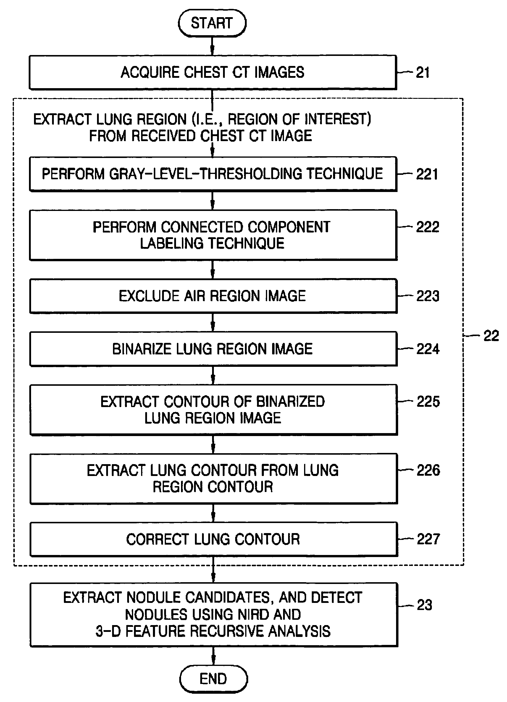
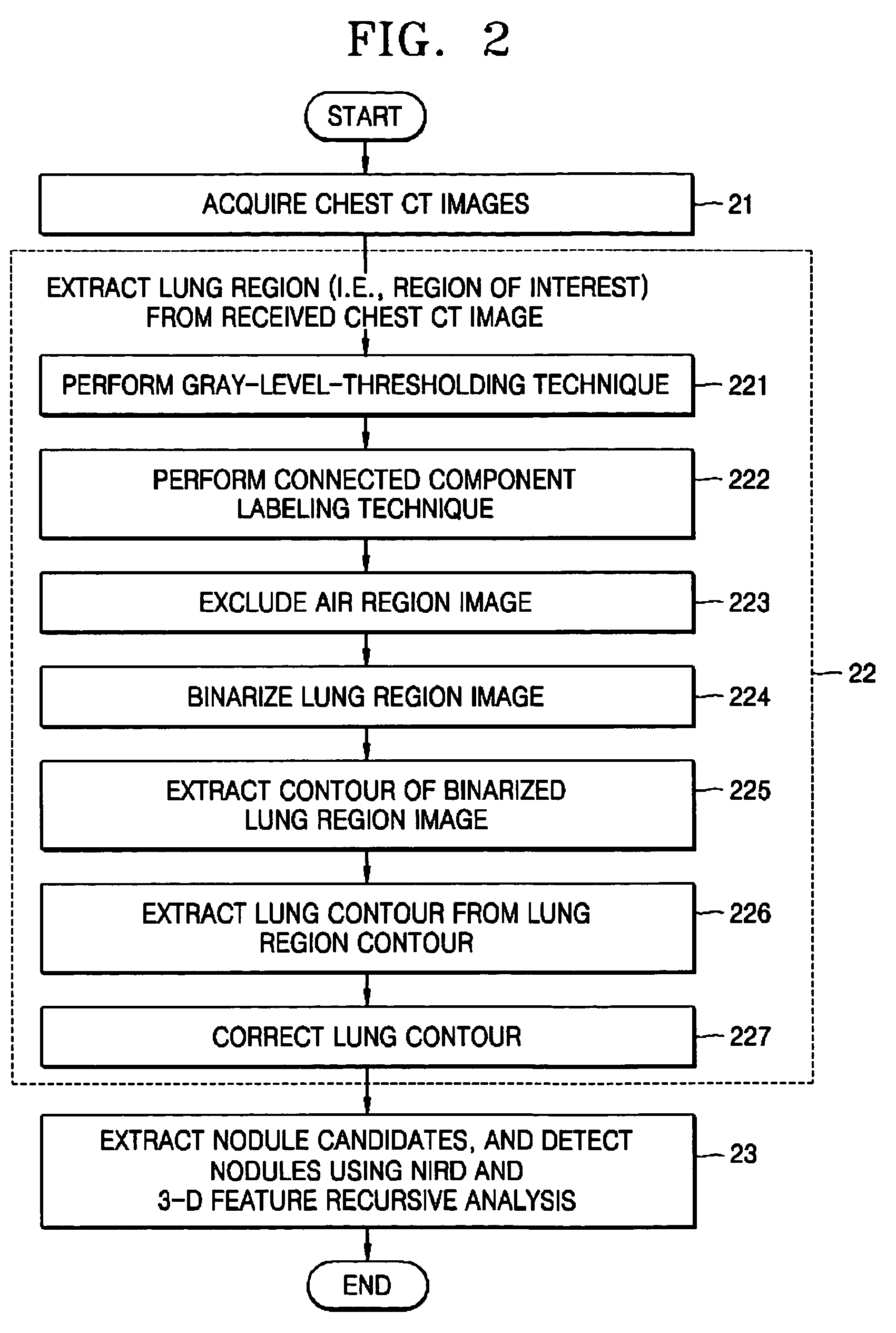
[0031] The present invention will now be described more fully with reference to the accompanying drawings, in which exemplary embodiments of the invention are shown. The invention may, however, be embodied in many different forms and should not be construed as being limited to the embodiments set forth herein; rather, these embodiments are provided so that this disclosure will be thorough and complete, and will fully convey the concept of the invention to those skilled in the art. In the drawings, the forms of elements are exaggerated for clarity. To facilitate understanding, identical reference numerals have been used, where possible, to designate identical elements that are common to the figures.

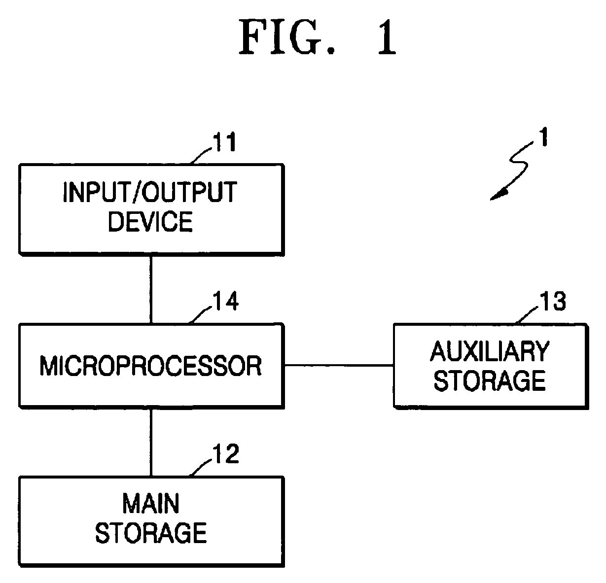
[0032]FIG. 1 is a block diagram of a hardware system 1 to which the present invention is applied. Referring to FIG. 1, the hardware system 1 includes an input / output device 11, main and auxiliary storages 12 and 13, and a microprocessor 14. The input / output device 11 is used by an externa...


PUM


Abstract

A method of automatically detecting pulmonary nodules is provided, including the operations of acquiring a chest computed tomography (CT) image, extracting a lung region from the chest CT image, extracting a group of nodule candidates from the lung region using a gray-level thresholding technique and a three-dimensional (3-D) region growing technique, and performing 3-D feature recursive analysis on all of the nodule candidates.

Description

BACKGROUND OF THE INVENTION [0001] This application claims the benefit of Korean Patent Application No. 2003-64722, filed on Sep. 18, 2003, in the Korean Intellectual Property Office, the disclosure of which is incorporated herein in its entirety by reference. [0002] 1. Field of the Invention [0003] The present invention relates to a method of automatically detecting pulmonary nodules from a chest computed tomographic (CT) image, and more particularly, to a method of automatically detecting pulmonary nodules using three-dimensional feature-analysis techniques and a computer readable recording medium which stores a program for executing the method. [0004] 2. Description of the Related Art [0005] Pulmonary masses or pulmonary nodules are represented as circular shades enclosed by distinctive boundaries on chest radiographs. Shades with diameters of 30 mm or less are classified as pulmonary nodules, and shades with diameters of more than 30 mm are classified as pulmonary masses. A sing...

Claims


Application Information

Patent Timeline
IPC IPC(8): G06F19/00G06T7/00G06V10/26
CPCG06K9/34G06T2207/30061G06T7/0012G06K2209/05G06V10/26G06V2201/03
Inventor LEE, JEONG WONKIM, SEUNGHWANKIM, YOUN TAE
Owner ELECTRONICS & TELECOMM RES INST
Why Eureka
Try Eureka