High resolution X ray, gamma ray and electron ray microscope

An electron ray and high-resolution technology, which is applied in the field of microscope systems, can solve problems such as the inability to realize large-angle refraction and the inability to form a microscopic magnification system, and achieve high resolution and improve the resolution limit.

Inactive Publication Date: 2016-08-31
顾士平
View PDF7 Cites 7 Cited by
  • Summary
  • Abstract
  • Description
  • Claims
  • Application Information

AI Technical Summary

Problems solved by technology

[0004] X-rays and γ-rays cannot form a microscopic magnification system because they cannot achieve large-angle refraction

Method used


Image

  • High resolution X ray, gamma ray and electron ray microscope
  • High resolution X ray, gamma ray and electron ray microscope

Examples


example 1

[0051] Preferred Example 1: Principle Block Diagram

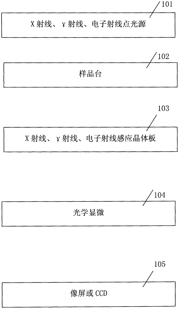
[0052] Such as figure 1 Shown, X-ray, gamma ray, electron ray microscope is by X-ray, gamma ray, electron ray point light source (101); Sample stage (102); X ray, gamma ray, electron ray crystal induction plate (103); micro-magnification system (104); composed of image screen or CCD (105);

[0053] X-ray, gamma ray, electron ray point light source (101), "point light source" passes through the sample on the sample stage (102), forms enlarged transmission image on X-ray, gamma ray, electron ray crystal induction plate (103); X X-ray, gamma ray, electron ray crystal induction plate (103), the crystal induction plate (103) converts X-ray, gamma ray, electron ray image into visible light image; the visible light image is amplified by optical microscope magnification system (104) again Display on image screen or CCD (105);

[0054] There are two sets of enlarged imaging systems in the present invention: (1) by X-ray, γ-ray, e...

example 2

[0059] Preferred Example 2: System Resolution Analysis:

[0060] Due to the diffraction characteristics of waves, according to the Abbe imaging principle, the imaging resolution of waves is:

[0061] δ=0.61λ / (n*sinα)

[0062] λ is the wavelength of illumination light; n is the refractive index; α lens numerical aperture;

[0063] The wavelength of X-rays is 0.01A-100A, and the wavelength of gamma rays is electromagnetic waves less than 0.01A wavelength;

[0064] Due to the diffraction of waves, there will be diffraction spots; it will affect the further improvement of the resolution of the existing optical micromagnification system mirror and electron microscope;

[0065] Method 1 uses X-rays and γ-rays with shorter wavelengths, thereby greatly improving the resolution limit of the microscope;

[0066] Method 2 uses electron rays with higher energy and shorter wavelength, thereby greatly improving the resolution limit of the microscope.

example 3

[0067] Preferred Example 3: Working Principle

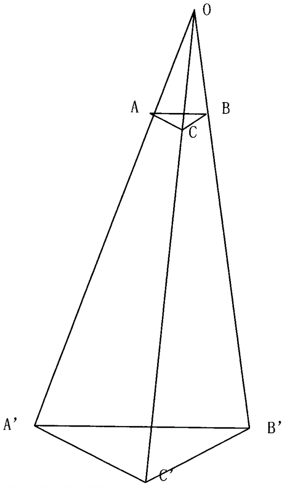
[0068] Resolution X-ray, γ-ray, and electron ray microscopes consist of two sets of magnification systems; the first magnification system uses X-ray, γ-ray, and electron ray point sources to geometrically magnify the object and display it on the imaging plate; the second set The magnification system enlarges the image on the imaging board; the magnification of the whole system is the magnification of the first set of magnification system × the magnification of the second set of magnification system;

[0069] The first magnification system, method 1, uses a point light source to magnify the sample by using the characteristics of the linear propagation of light; method 2: uses the electronic lens magnification system to magnify the sample;

[0070] The crystal sensing plate (103) converts the X-ray image into a visible light image;

[0071] The crystal sensing plate (103) converts the γ-ray image into a visible light image;

[00...


PUM


Abstract

The invention relates to a high resolution X ray, gamma ray and electron ray microscope. The microscope is composed of two sets of amplifying systems. A method 1 comprises that a first amplifying system adopts rectilinear propagation of wave, amplifies an object by utilizing the X ray, gamma ray and electron ray and carried out imaging on a crystal induction plate; a method 2 comprises that an existing transmission-type electron microscope system is utilized for amplifying the object and imaging on the crystal induction plate; the crystal induction plate converts images of the X ray, gamma ray and electron ray into visible light images; a second amplifying system amplifies the visible light images on an imaging plate; amplification factor of the whole system is amplification factor of the first amplifying system*amplification factor of the second amplifying system; and a two-stage amplification system is utilized, so that resolution higher than that of an existing transmission electron microscope is realized.

Description

1. Technical field [0001] This patent relates to a microscope using X-rays, γ-rays, and electron ray imaging, and in particular to a microscope system using secondary imaging. 2. Background technology [0002] Now there are optical transmission microscope, optical reflection microscope, electron transmission microscope; [0003] Due to the diffraction of light, the resolution limit of the visible light optical microscope magnification system mirror is 0.2 microns; the resolution limit of the electron transmission microscope is 0.02 nanometers; [0004] X-rays and γ-rays cannot form a microscopic magnification system because they cannot achieve large-angle refraction. 3. Contents of the invention [0005] Problem to be solved: [0006] Address the resolution limit of existing microscopes. [0007] Technical solutions: [0008] X-ray, γ-ray, electron ray microscope consists of X-ray, γ-ray, electron ray point light source (101); sample stage (102); X-ray, γ-ray, electron...

Claims


Application Information

Patent Timeline
Patent Type & Authority Applications(China)
IPC IPC(8): G02B21/00G01N23/04
CPCG02B21/00G01N23/04
Inventor 顾士平顾海涛崔崇
Owner 顾士平
Why Eureka
Try Eureka