A preparation method of a large-area thin-area transmission electron microscope sample

A technology for transmission electron microscope samples and electron microscope samples, which is applied in the field of preparation of large-area thin-area samples, can solve the problems of discontinuous thin areas of samples, expensive equipment, damage, etc., and achieve the effect of eliminating incomplete surface of samples and controllable areas

Active Publication Date: 2017-03-29
INST OF GEOLOGY & GEOPHYSICS CHINESE ACAD OF SCI
View PDF5 Cites 2 Cited by
  • Summary
  • Abstract
  • Description
  • Claims
  • Application Information

AI Technical Summary

Problems solved by technology

In the current technology, researchers at home and abroad mainly use two methods to prepare TEM samples. One is the focused ion beam-scanning electron microscope dual-beam system (FIB-SEM), which has been widely used in recent years. This method is generally used for Fixed-point cutting of samples, the size is usually 15×5μm, the thin area available for observation is too small, and the equipment is expensive and expensive. In addition, the damage of the Ga ion beam used for cutting to the sample is also a major factor restricting its wide application; Another method is the traditional ion thinning method. Generally, a sample with a diameter of 3 mm is cut out, and the surface is ground to less than 100 μm. thin area
However, this method is only suitable for ordinary homogeneous material samples, but many composite materials, especially minerals, meteorites, fossils and other samples in the geological field, require a large thin area to determine the complex microstructure, composition and composition of minerals. and the thin area of ​​the sample made by this ion thinning method is discontinuous, and the width of the thin area is generally less than 20 μm, which is far from meeting the requirements of transmission electron microscopy analysis of many solid samples; in addition, with the nanometer ion probe With the development of in-situ techniques such as needles and electron probes, there is an urgent need for a sample preparation method that can be used for these in-situ techniques and for transmission electron microscopy analysis.
Therefore, these problems existing in the current technology have greatly restricted the expansion and application of transmission electron microscopy testing technology.

Method used


Image

  • A preparation method of a large-area thin-area transmission electron microscope sample
  • A preparation method of a large-area thin-area transmission electron microscope sample
  • A preparation method of a large-area thin-area transmission electron microscope sample

Examples


Embodiment Construction

[0023] Further description will be given below through examples and in conjunction with the accompanying drawings.

[0024] According to this embodiment Figure 4 The preparation process shown is for the preparation of large-area transmission electron microscope samples, and the specific steps include:

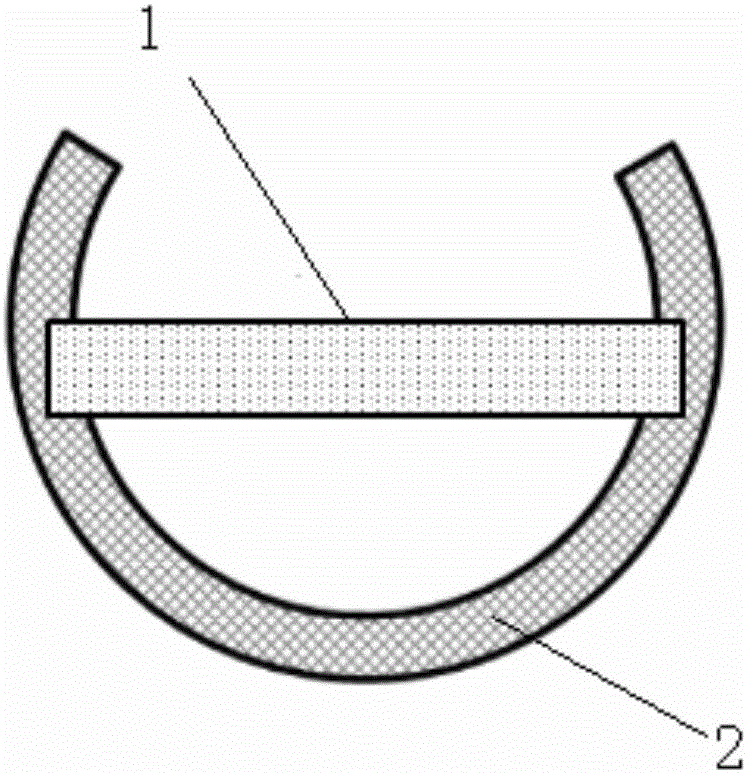
[0025] (1) Cutting and processing of the sample: first cut the material sample to be tested into strips of about 2.8×1×1mm in length×width×height, stick them on the glass slide with paraffin, put them in the fixture and use the particle size of 30μm in turn. , 10μm, 5μm, and 1μm sandpaper to polish and grind the sample to less than 200μm, and glue the ground sample to the semicircular copper ring with AB, such as figure 1 shown;

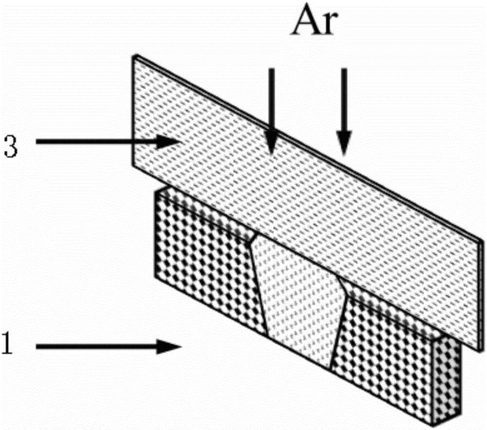
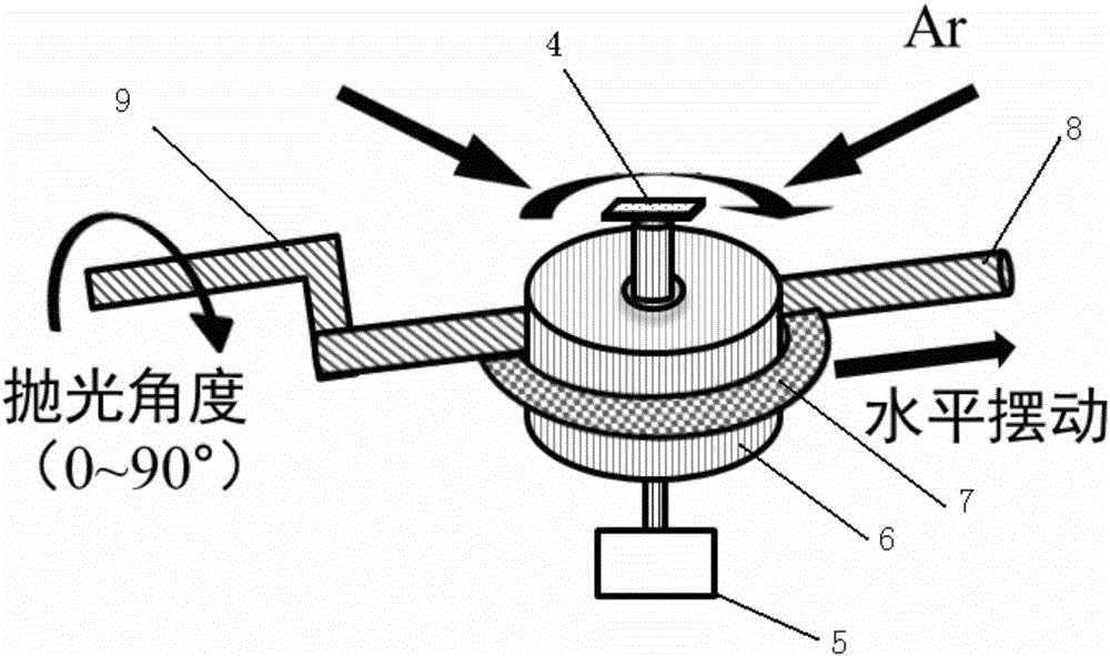
[0026] (2) The sample is ion-cut: put the copper ring with the sample on the sample stage of the ion slicer, and the pre-vacuum degree is better than 1×10 -4 Pa, so that the argon ion beam and the sample plane are at zero degrees for ion cutting, ...


PUM


Abstract

The invention relates to a method for preparing a large-area transmission electron microscope sample. The sample is cut and ground into a strip and then ion cutting is carried out by an argon ion beam so as to enable a thickness of a target region to reach 10mum; then, the sample is transferred to an ion beam polissoir for flat placement, a sample table is adjustable in angle and can rotate and swing, the sample is subjected to polishing and thinning by the argon ion beam sequentially at angles of 18 degrees, 12 degrees, 9 degrees, 6 degrees and 3 degrees, and variations of the sample can be observed by a stereoscope; finally, the sample is cleaned by a low-voltage ion beam, an amorphous layer generated in the sample preparing process is avoided, and the high-quality transmission electron microscope sample with a large area of thin region can be obtained, so that the high-quality transmission electron microscope sample with the large area of thin region can be applied to transmission electron microscope testing of complex materials such as minerals, meteorolite and the like and an in situ analysis technology combining a nano particle probe / electronic probe and the like.

Description

technical field [0001] The invention belongs to the technical field of electron microscope testing of solid samples such as minerals and meteorites, and relates to a method for preparing large-area thin-area samples suitable for transmission electron microscopy. Background technique [0002] At present, in the field of electron microscopy testing, due to the limitation of the penetration ability of the electron beam, in order to obtain better transmission electron microscopy results, it is necessary to obtain high-quality samples. This sample needs to have a thin area covering the target area, the thickness Generally around 100nm, if the sample is observed with a high-resolution image, the thickness of the sample is required to be less than 50nm. In the current technology, researchers at home and abroad mainly use two methods to prepare TEM samples. One is the focused ion beam-scanning electron microscope dual-beam system (FIB-SEM), which has been widely used in recent years...

Claims


Application Information

Patent Timeline
Patent Type & Authority Patents(China)
IPC IPC(8): G01N1/28
CPCG01N1/286G01N1/32G01N23/2202G01N23/225G01N2001/2866G01N2001/2873
Inventor 谷立新赵旭晁林杨挺
Owner INST OF GEOLOGY & GEOPHYSICS CHINESE ACAD OF SCI
Why Eureka
Try Eureka