Auxiliary cancer diagnosis method based on digital pathological images
A technology for digital pathology and auxiliary diagnosis, applied in the field of auxiliary diagnosis of cancer based on digital pathological images, which can solve the problems of insufficient consistent ratio and inability to provide diagnosis basis to support diagnosis results.
- Summary
- Abstract
- Description
- Claims
- Application Information
AI Technical Summary
Problems solved by technology
Method used
Image

Examples
Embodiment Construction
[0020] The present invention will be further described below in conjunction with the accompanying drawings and specific embodiments.
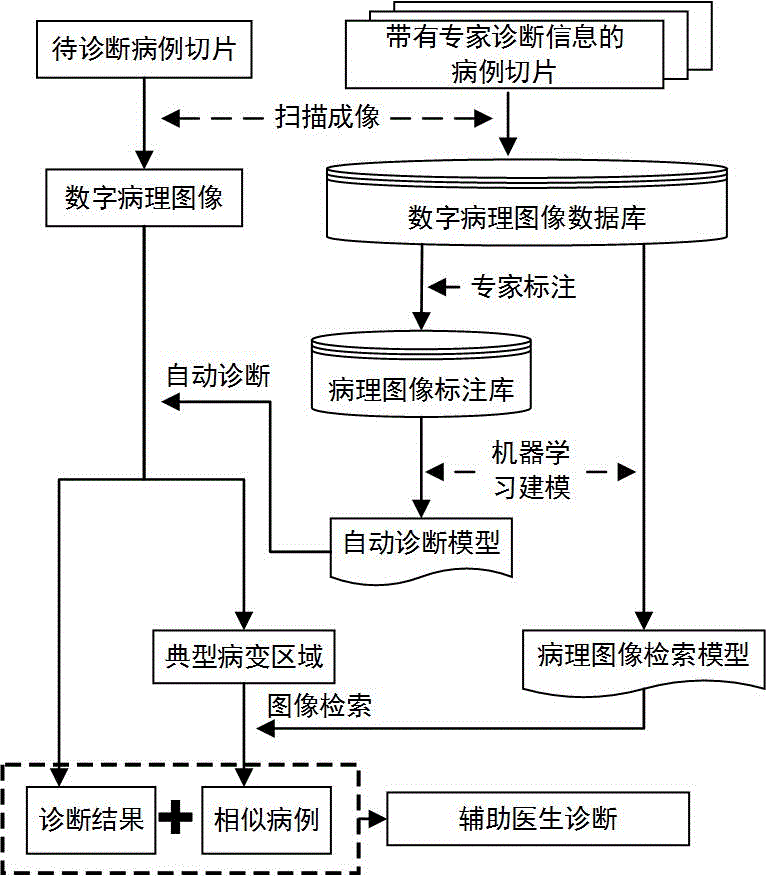
[0021] Such as figure 1 As shown, a cancer-aided diagnosis method based on digital pathological images includes the following specific steps:
[0022] Step 1: Use a slice scanner to scan the digital pathological slice into the electronic calculation, and store it as a digital image matrix with RGB three-channel
[0023] In order to meet the needs of cancer diagnosis, pathological slides are generally scanned under a 40X mirror, which makes the scale of the entire digital pathological slide reach 90,000×90,000 pixels. In order to facilitate reading, transmission and processing, digital pathological slides are generally divided into blocks stored in the form.
[0024] Step 2: Screen the digital pathological slices with diagnostic information of pathology experts to establish a digital pathological image database, and select some typical digital...
PUM
 Abstract
Description
Claims
Application Information