Automatic extraction method of human chest organ tissue

An automatic extraction and chest technology, which is applied in the field of medical image processing, can solve the problems that the image segmentation technology is not clear enough, the boundaries of organs and tissues are not smooth, and affect the observation of researchers and clinicians, and achieve smooth boundaries, clear image segmentation, and smooth volume Effect

Active Publication Date: 2017-01-25
HEILONGJIANG TUOMENG TECH CO LTD
View PDF1 Cites 6 Cited by
  • Summary
  • Abstract
  • Description
  • Claims
  • Application Information

AI Technical Summary

Problems solved by technology

[0003] In order to solve the problem that the existing image segmentation technology is not clear enough, the boundary of organs and tissues is not smooth, which affects the observation of scientific researchers and clinicians, and further provides an automatic extraction method of human chest organs and tissues

Method used


Image

  • Automatic extraction method of human chest organ tissue
  • Automatic extraction method of human chest organ tissue
  • Automatic extraction method of human chest organ tissue

Examples


specific Embodiment approach 1

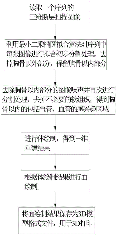
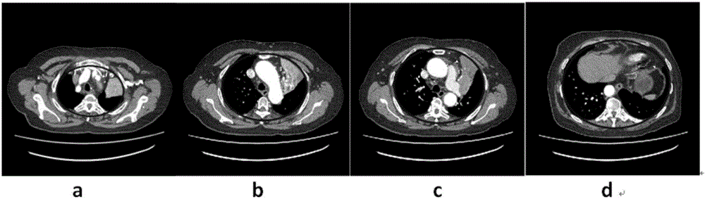
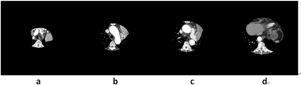
[0043] Specific implementation mode one: see figure 1 , Figure 2 to Figure 7 Describe this embodiment, such as Figure 2 to Figure 7 Shown: figure 2 is the original CT image; image 3 is the result after applying the least squares ellipse fitting algorithm to segment; Figure 4 It is the result of applying the 3D TV-L1 algorithm to denoise and smooth; Figure 5 It is the result of secondary segmentation by applying 3D TV-L1 algorithm; Figure 6 Render the result for the 3D volume; Figure 7 Plot the results for 3D faces. The realization process of the automatic extraction method of human chest organ tissue described in this embodiment is:

[0044] Step 1, reading a sequence of three-dimensional tomographic images;

[0045] Step 2. Use the least squares ellipse fitting algorithm to perform a preliminary segmentation process on each image in the sequence, remove the part outside the sternum, and keep the part inside the sternum; applying the ellipse fitting method to t...

specific Embodiment approach 2

[0080] Embodiment 2: In this embodiment, after the volume rendering in step 4, manual interactive segmentation can be performed, and unnecessary regions can be manually selected for deletion. Other steps are the same as in the first embodiment.

specific Embodiment approach 3

[0081] Embodiment 3: In this embodiment, in step 5, the MC (marching cube) algorithm is used for surface rendering to obtain a three-dimensional reconstruction result. Other steps are the same as those in Embodiment 1 or 2.


PUM


Abstract

The invention discloses an automatic extraction method of human chest organ tissue, belongs to the technical field of medical image processing, and aims to solve the problems that boundaries of organ tissue are unsmooth and observation by researchers and clinicians is affected due to the fact that an existing image segmentation technology is not clear enough. The automatic extraction method is characterized by comprising steps as follows: reading three-dimensional tomography images; performing fitting and primary segmentation processing on each image in a sequence with a least square ellipse fitting algorithm, parts outside sternums are removed, and parts inside the sternums are reserved; image noise of the parts inside the sternums is removed, segmentation processing is performed again to remove unnecessary soft tissue, and regions of interest, including trachea and blood vessels, inside the sternums are obtained; volume rendering and surface rendering are performed. Medical image segmentation has great significance in three-dimensional visualization, three-dimensional positioning, operation planning formulation and computer assisted diagnosis. With the adoption of the automatic segmentation method, the effect of subjective factors of an observer is completely avoided, the data processing speed is increased, and the repeatability is good.

Description

technical field [0001] The invention relates to an automatic extraction method of human chest organ tissue, which belongs to the technical field of medical image processing. Background technique [0002] In the past 20 years, with the rapid development of medical imaging equipment, various medical institutions have to generate a large amount of medical imaging data every day. Computed tomography (CT), magnetic resonance imaging (magnetic resonance imaging, MRI) and other imaging technologies have become observation organs. important means of organization. The results of medical image processing enable researchers and clinicians to intuitively and clearly observe the normal and lesion positions inside the human body. Therefore, relevant experts at home and abroad have always attached great importance to medical image processing technology, and medical image segmentation is a classic problem in image processing technology. The existing image segmentation technology is not cl...

Claims


Application Information

Patent Timeline
Patent Type & Authority Applications(China)
IPC IPC(8): G06T7/11G06T5/00G06F19/00G06T15/08G06T17/00
CPCG06T5/002G06T15/08G06T17/00G06T2207/10081G06T2207/10088G06T2207/30061G06T2207/30096G06T2207/30101G06T2210/41
Inventor 邱兆文端金鸣
Owner HEILONGJIANG TUOMENG TECH CO LTD
Why Eureka
Try Eureka