Fundus image retinal vessel segmentation method and system based on deep learning

A technology for retinal blood vessels and fundus images, which can be used in image analysis, image enhancement, image data processing, etc., and can solve problems such as limited feature extraction.

Active Publication Date: 2017-02-15
SOUTH CHINA UNIV OF TECH
View PDF3 Cites 213 Cited by
  • Summary
  • Abstract
  • Description
  • Claims
  • Application Information

AI Technical Summary

Problems solved by technology

The main difficulty of this method lies in feature extraction and image segmentation. For machine learning methods, feature engineering is very important. Traditional methods mainly use Gabor filtering and other methods, and the extraction features are limited. In recent years, with the development of deep learning, using deep learning The feature extraction of images has a good effect, and some people try to use deep learning for blood vessel segmentation

Method used


Image

  • Fundus image retinal vessel segmentation method and system based on deep learning
  • Fundus image retinal vessel segmentation method and system based on deep learning
  • Fundus image retinal vessel segmentation method and system based on deep learning

Examples


Embodiment

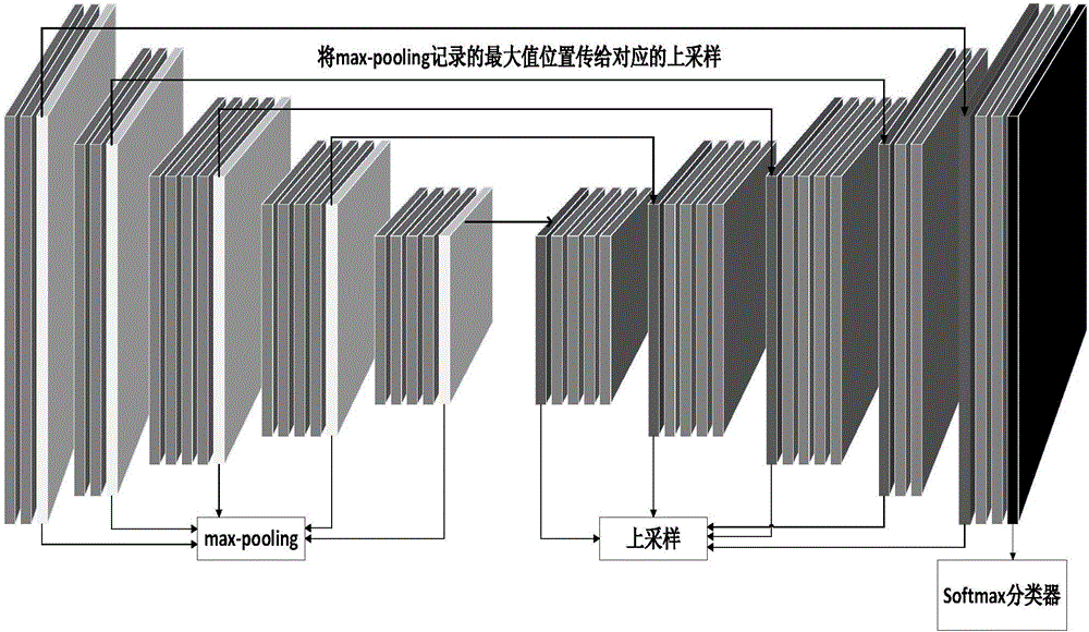
[0080] The retinal blood vessel segmentation method of the fundus image based on deep learning of the present invention is as follows: Figure 4 shown, including the following steps:

[0081] Step 1: Preprocessing the fundus images in the dataset;

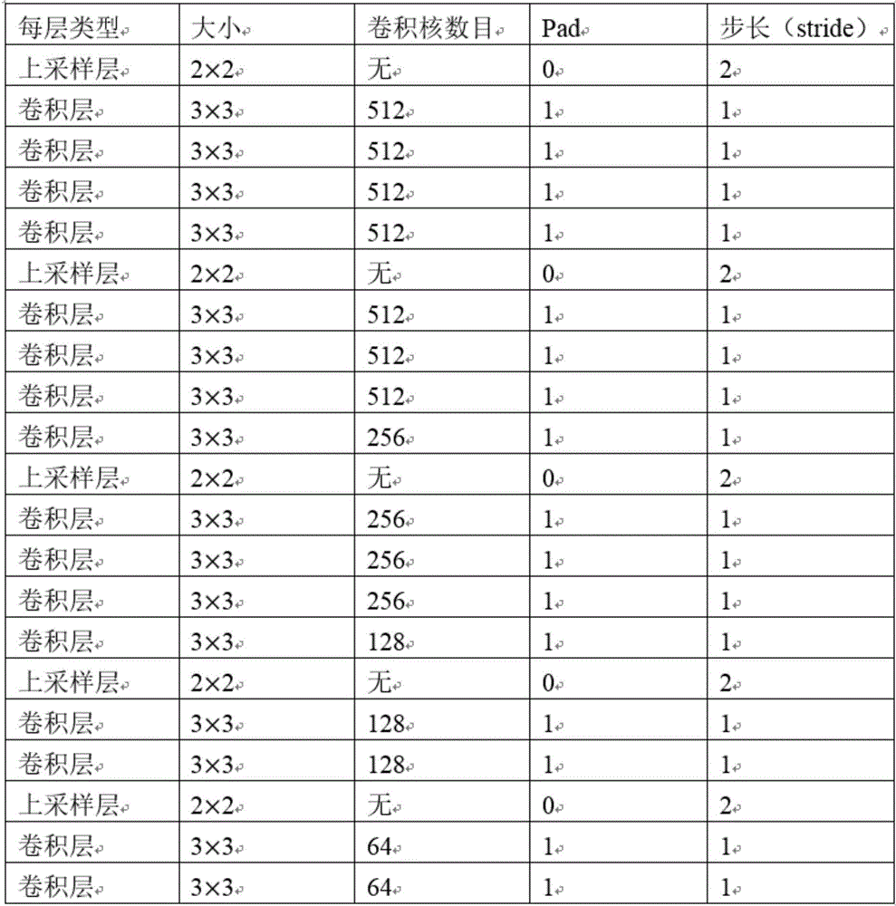
[0082] Step 2: Train the convolutional neural network with training samples;

[0083] Step 3: Extract the last layer of convolution output features from the trained convolutional neural network to train a random forest classifier;

[0084] Step 4: Fuse the pixel classification results of the convolutional neural network with the results of the random forest classifier;

[0085] Step 5: Use the trained convolutional neural network model to segment the test sample to obtain the final segmentation result.

[0086] Specifically, in step 1, the fundus images in the data set are preprocessed, and the fundus images in the data set are divided into training samples and test samples. The blood vessels and non-vessels in the original ima...


PUM


Abstract

The invention discloses a fundus image retinal vessel segmentation method and a fundus image retinal vessel segmentation system based on deep learning. The fundus image retinal vessel segmentation method comprises the steps of performing data amplification on a training set, enhancing an image, training a convolutional neural network by using the training set, segmenting the image by using a convolutional neural network segmentation model to obtain a segmentation result, training a random forest classifier by using features of the convolutional neural network, extracting a last layer of convolutional layer output from the convolutional neural network, using the convolutional layer output as input of the random forest classifier for pixel classification to obtain another segmentation result, and fusing the two segmentation results to obtain a final segmentation image. Compared with the traditional vessel segmentation method, the fundus image retinal vessel segmentation method uses the deep convolutional neural network for feature extraction, the extracted features are more sufficient, and the segmentation precision and efficiency are higher.

Description

technical field [0001] The invention relates to the fields of machine learning and image processing, and is aimed at the research of medical image semantic segmentation technology, especially a method and system for retinal blood vessel segmentation of fundus images based on deep learning. Background technique [0002] In recent years, with the development of image processing technology, image segmentation technology has begun to be applied to the field of fundus image segmentation. At present, many researchers at home and abroad have proposed a variety of retinal blood vessel segmentation algorithms for fundus images, which are mainly divided into the following: Directions: methods based on vessel tracking, methods based on matched filtering, methods based on deformation models, and methods based on machine learning. [0003] The method based on matched filtering is to convolve the filter with the image to extract the target object. Since the gray level of the retinal vesse...

Claims


Application Information

Patent Timeline
Patent Type & Authority Applications(China)
IPC IPC(8): G06T7/00G06T7/11
CPCG06T7/0012G06T2207/20032G06T2207/20081G06T2207/30101
Inventor 余志文马帅吴斯纪秋佳韩国强
Owner SOUTH CHINA UNIV OF TECH
Why Eureka
Try Eureka