Anal levator shaft plane image processing method and device thereof

An image processing and axial plane technology, applied in the field of medical ultrasound imaging, can solve the problems of adjusting the position and size of the sampling frame, the accuracy of the sagittal plane is difficult to ensure, and it is difficult to locate the position of the posterior and inferior border of the pubic symphysis, so as to improve the accuracy. and efficiency effects

Active Publication Date: 2017-03-08
SONOSCAPE MEDICAL CORP
View PDF1 Cites 6 Cited by
  • Summary
  • Abstract
  • Description
  • Claims
  • Application Information

AI Technical Summary

Problems solved by technology

In the actual processing process, due to differences in doctors' techniques, the accuracy of the mid-sagittal plane of the pelvic floor is difficult to guarantee, and due to the lack of understanding of three-dimensional space, it is difficult to locate the position of the posterior inferior edge of the pubic symphysis in the separate volume data to adjust the sampling Box position and size
Therefore, it is difficult for most doctors to efficiently obtain plane images of the levator ani muscle axis in 3D, which reduces the diagnostic accuracy and efficiency

Method used


Image

  • Anal levator shaft plane image processing method and device thereof
  • Anal levator shaft plane image processing method and device thereof
  • Anal levator shaft plane image processing method and device thereof

Examples


Embodiment Construction

[0059] In order to enable those skilled in the art to better understand the solution of the present invention, the present invention will be further described in detail below in conjunction with the accompanying drawings and specific embodiments. Apparently, the described embodiments are only some of the embodiments of the present invention, but not all of them. Based on the embodiments of the present invention, all other embodiments obtained by persons of ordinary skill in the art without making creative efforts belong to the protection scope of the present invention.

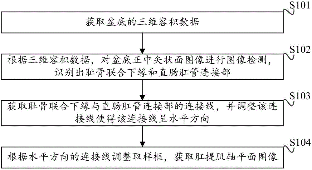
[0060] A flow chart of a specific embodiment of the levator ani axis plane image processing method provided by the present invention is as follows figure 1 As shown, the method includes:

[0061] Step S101: Acquiring three-dimensional volume data of the pelvic floor;

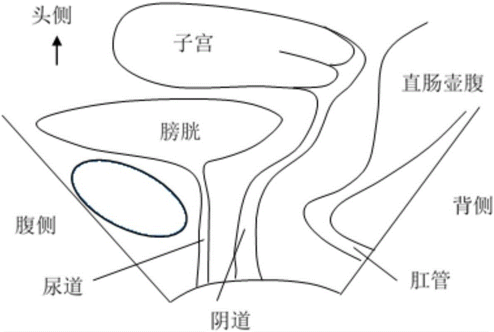
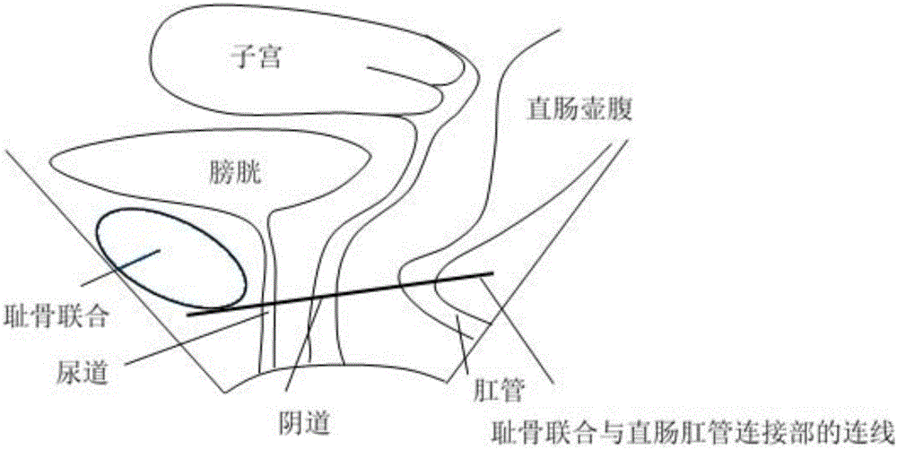
[0062] The mid-sagittal plane of the pelvic floor includes the pubic symphysis, urethra, bladder, vagina, uterus, rectal ampulla, anal canal,...


PUM


Abstract

The invention discloses an anal levator shaft plane image processing method and a device thereof. The method is characterized by acquiring three-dimensional volume data of a basin floor; according to the three-dimensional volume data, carrying out image detection on a basin-floor-center sagittal image and identifying a pubic symphysis lower margin and a rectum and anal canal connection portion; acquiring a connection line of the pubic symphysis lower margin and the rectum and anal canal connection portion, adjusting the connection line and making the connection line be in a horizontal direction; and according to the connection line of the horizontal direction, adjusting a sampling frame and acquiring an anal levator shaft plane image. By using the anal levator shaft plane image processing method and the device thereof, the pubic symphysis and the rectum and anal canal connection portion can be automatically identified so that the sampling frame is rapidly and accurately adjusted and an anal levator area image is acquired; and accuracy and efficiency of image processing are increased.

Description

technical field [0001] The invention relates to the technical field of medical ultrasonic imaging, in particular to a method and device for processing a plane image of a levator ani muscle axis. Background technique [0002] The levator ani muscle is composed of visceral pubone muscle, puborectalis muscle, and iliococcygeus muscle, and is the most important pelvic floor muscle supporting the pelvic organs. The levator ani muscle on both sides and the lower edge of the pubic symphysis jointly form the levator ani muscle hiatus, through which the urethra, vagina, and rectum pass. If the levator ani muscle is damaged, it will affect the support of the pelvic organs and cause prolapse. The axial plane of the levator ani muscle can well display the levator ani muscle hiatus and surrounding muscle groups, and evaluate the injury of the levator ani muscle. [0003] In pelvic floor ultrasound imaging, the biggest advantage of 3D / 4D ultrasound is that it can display the axis plane o...

Claims


Application Information

Patent Timeline
Patent Type & Authority Applications(China)
IPC IPC(8): G06T7/00
CPCG06T7/0012G06T2207/10136
Inventor 唐艳红孙慧李萍刘旭江艾金钦
Owner SONOSCAPE MEDICAL CORP
Why Eureka
Try Eureka