Three-dimensional image segmentation system and segmentation method thereof

A three-dimensional image and image technology, applied in the field of image processing, can solve the problems of sensitivity to noise and grayscale diversity, large amount of calculation, discontinuity, etc., to achieve the effect of short segmentation time, elimination of image noise, and smooth image edges.

Active Publication Date: 2017-03-08
SHENZHEN INST OF ADVANCED TECH CHINESE ACAD OF SCI
View PDF1 Cites 4 Cited by
  • Summary
  • Abstract
  • Description
  • Claims
  • Application Information

AI Technical Summary

Problems solved by technology

At present, there are many image segmentation algorithms, but none of them can be applied to all kinds of medical images, and the accuracy of many algorithms applied to 3D medical images is relatively low, and the program running time is relatively long, which cannot meet the real-time requirements of 3D medical images. sex analysis needs
[0003] Currently commonly used image segmentation methods have the following disadvantages: (1) Region-based segmentation methods are sensitive to noise and gray-level diversity, rely too much on the selection of seed points, and have a large amount of calculation.
(2) Edge detection methods mainly include serial and parallel methods: the segmentation result of the serial method is more dependent on the initial edge points, and inappropriate initial edge points may lead to wrong edges; the parallel method is sensitive to noise, and when the image of the edge You may get discontinuities or spurious boundaries with small changes in prime values
(3) The active contour model has the disadvantages that it is difficult to capture the concave boundary of the target and is sensitive to the initial contour line; the data of each pixel in the fuzzy C-means algorithm is independent of each other, and the spatial information of the image is not used; the genetic algorithm is good at global search, and the local Insufficient search ability, sensitive to noise

Method used


Image

  • Three-dimensional image segmentation system and segmentation method thereof
  • Three-dimensional image segmentation system and segmentation method thereof
  • Three-dimensional image segmentation system and segmentation method thereof

Examples


Embodiment Construction

[0047] Hereinafter, embodiments of the present invention will be described in detail with reference to the accompanying drawings. This invention may, however, be embodied in many different forms and should not be construed as limited to the specific embodiments set forth herein. Rather, these embodiments are provided to explain the principles of the invention and its practical application, so that others skilled in the art can understand various embodiments of the invention and various modifications as are suited to particular intended uses. The same reference numerals may be used to refer to the same elements throughout the specification and drawings.

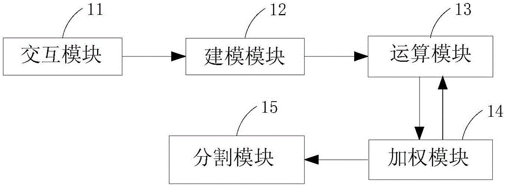
[0048] figure 1 It is a block diagram of a 3D image segmentation system in a preferred embodiment of the present invention.

[0049] refer to figure 1 , the 3D medical image segmentation system according to the embodiment of the present invention includes: an interaction module 11 , a modeling module 12 , an operation modul...


PUM


Abstract

The invention discloses a three-dimensional image segmentation system and a segmentation method thereof. The segmentation method comprises steps of: selecting an image with a frame so as to obtain a target image including a target object; according to the target image, establishing a foreground model and a background module; calculating undirected connected graphs and undirected weighted graphs of the foreground and the background; assigning weight value to the undirected connected graphs so as to obtain weighted undirected connected graphs; calculating a minimal cutest of the undirected weighted graphs; and segmenting the weighted undirected connected graphs according to the minimal cutest so as to obtain two unconnected sub graphs, thereby achieving segmentation of the foreground and the background of the target image. According to the invention, three-dimensional medial image data can be directly segmented; used time is short, segmentation precision is high, the robustness of the segmentation results is good and a cavity phenomenon will not be caused; and in addition, the visualization effect of the three-dimensional image segmentation system and the segmentation method thereof is good, image noise can be effectively eliminated and the edges of the segmented images are quite smooth.

Description

technical field [0001] The invention belongs to the technical field of image processing, and in particular relates to a three-dimensional image segmentation system and a segmentation method thereof. Background technique [0002] Medical image segmentation is an important part of medical image processing, and the segmentation results play a vital role in clinical diagnosis and treatment. The purpose of medical image segmentation is to separate anatomical structures of interest from medical images or to locate the location and shape of disease sources, which directly determines the accuracy of subsequent analysis. At present, there are many image segmentation algorithms, but none of them can be applied to all kinds of medical images, and the accuracy of many algorithms applied to 3D medical images is relatively low, and the program running time is relatively long, which cannot meet the real-time requirements of 3D medical images. gender analysis needs. [0003] Currently com...

Claims


Application Information

Patent Timeline
Patent Type & Authority Applications(China)
IPC IPC(8): G06T7/11
CPCG06T2207/10012G06T2207/30004
Inventor 余绍德姬治华陈璐明江帆伍世宾谢耀钦
Owner SHENZHEN INST OF ADVANCED TECH CHINESE ACAD OF SCI
Why Eureka
Try Eureka