Comprehensive analysis system for xerophthalmia
A comprehensive analysis and analysis system technology, applied in the field of dry eye detection equipment, can solve the problems of Keratograph 5M equipment bulky, long duration of tear secretion test method, incompatibility with ophthalmic examination equipment, etc., to achieve adjustable light source and brightness, reduce Check the effect of difficulty and convenience for doctors to operate
- Summary
- Abstract
- Description
- Claims
- Application Information
AI Technical Summary
Problems solved by technology
Method used
Image
Examples
Embodiment 1
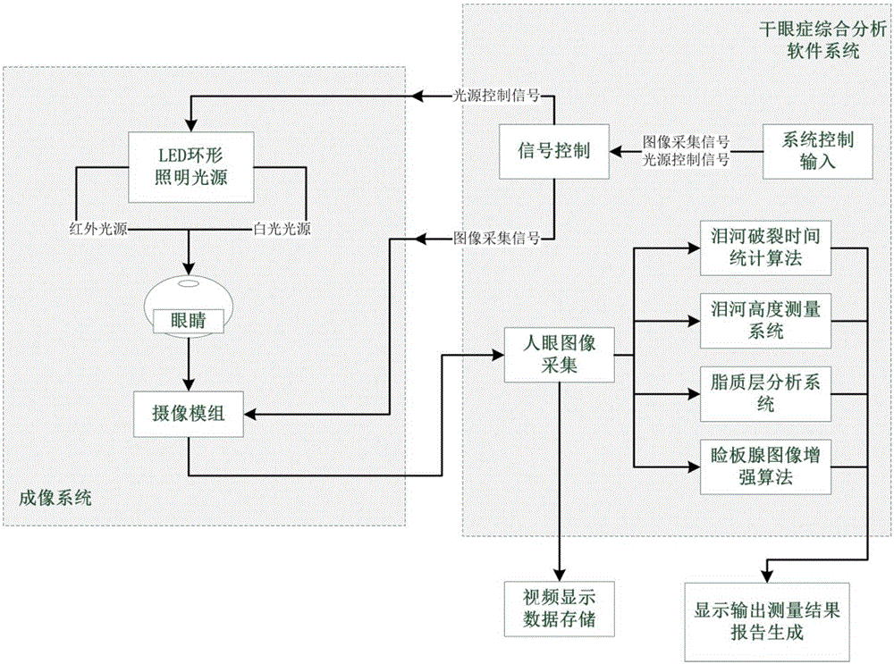
[0043] like figure 1 Shown is an implementation form according to the present invention, which includes an imaging system for photographing a patient's eye, an analysis system for performing multiple symptom analysis on the image, and a video terminal for displaying the image.
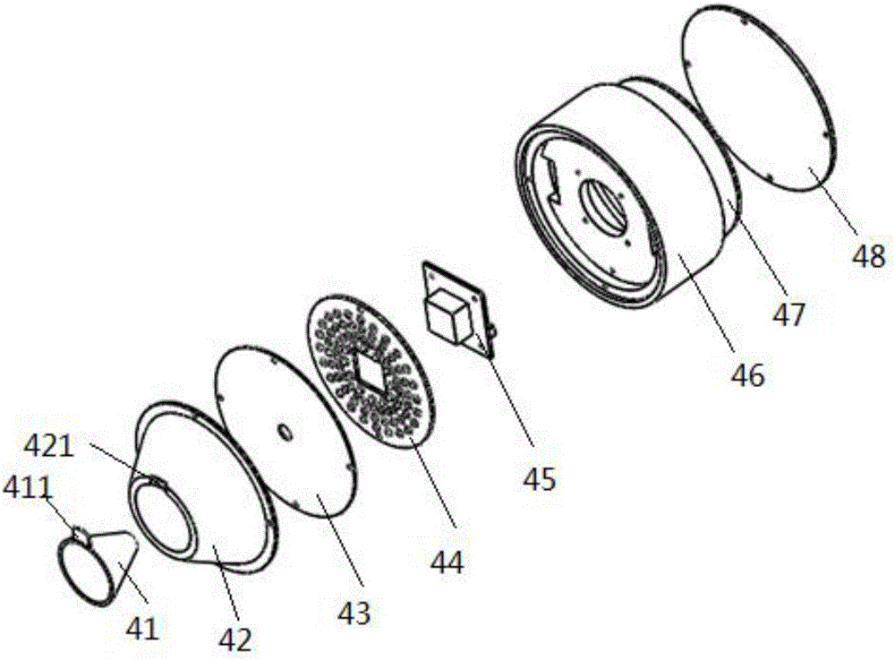
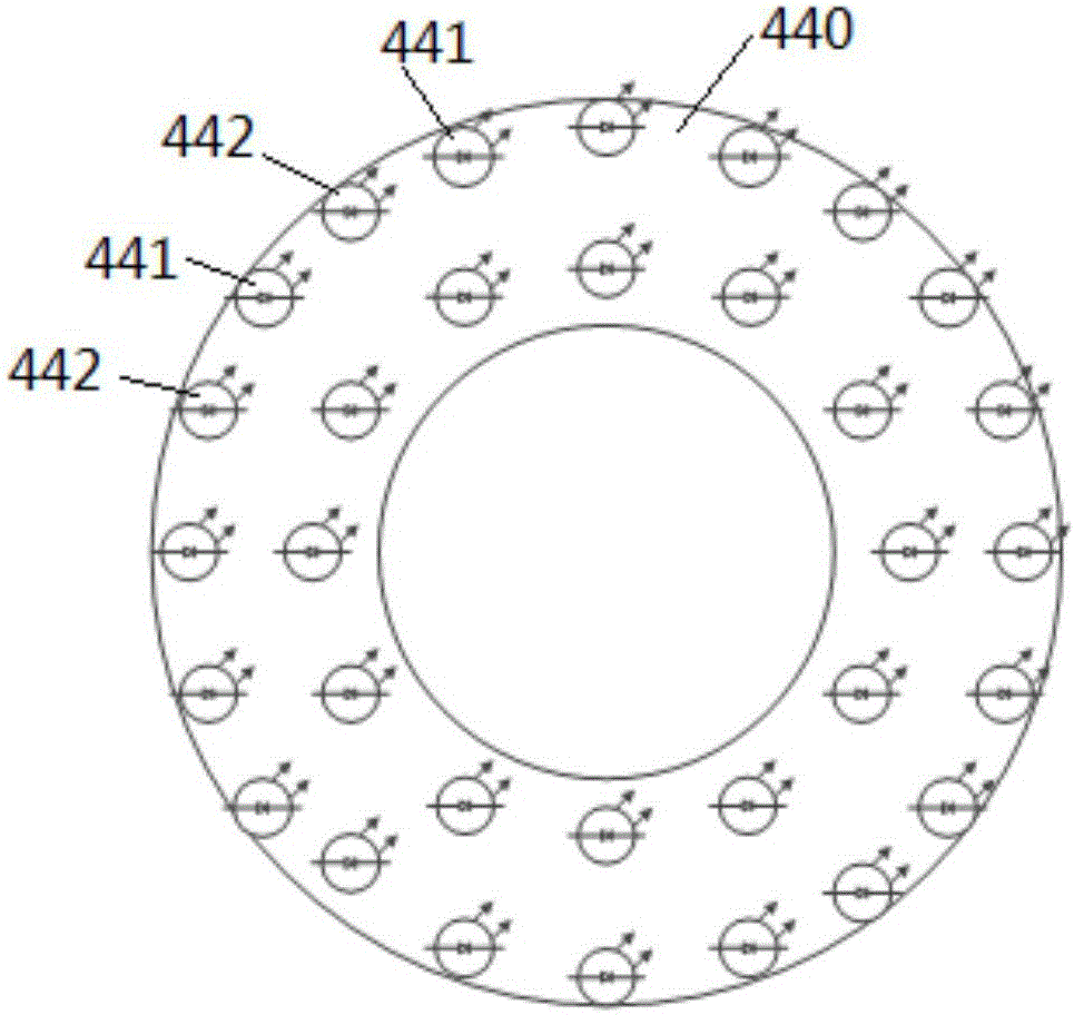
[0044] In the present invention, the imaging system is arranged in an integrated probe assembly 4, such as figure 2 As shown, the probe assembly includes an imaging assembly, an illumination light source 44, a camera module 45, and a control board 47 arranged in sequence. The imaging assembly, the illumination light source 44 and the camera module 45 are fixed on one side of the camera fixing device 46. The control board 47 is fixed on the other side of the camera fixing device 46, and at the same time, it is closed with a cover plate 48 to form a complete structure. The fixing sleeve 42 is configured as a light-concentrating cavity structure, the light-concentrating cavity is provided with openings ...
Embodiment 2
[0052] On the basis of Example 1, as figure 2 As shown, a baffle 411 is laterally provided at the flat bottom end of the conical cylinder 41, and a positioning slot 421 is opened on the small opening of the condensing cavity. In order to satisfy different detection functions, the imaging mode needs to be switched, so The conical cylinder 41 needs to be removed from the fixing sleeve. Therefore, in this embodiment, the conical cylinder 41 is detachably installed on the fixing sleeve 42, and the blocking piece 411 is clamped on the fixing sleeve 42. In the positioning slot 421, the Placido conical cylinder is designed to be removed during use to fulfill the needs of different functional imaging.
Embodiment 3
[0054] On the basis of Embodiment 1 or Embodiment 2, a diffuse reflection coating is coated on the inner side wall of the light-collecting cavity, and the light emitted from the illumination light source is uniformly reflected to the cone through the diffuse reflection coating. On the conical surface of the tube, the outgoing light is concentrated on the conical surface of the conical tube, which enhances the effectiveness of the irradiated light, reduces polarized light and light leakage, so that the outgoing light is concentrated on the patient's eyes, providing better lighting effects, and then Improve the imaging effect and improve the accuracy, objectivity and stability of the measurement results.
PUM
 Abstract
Description
Claims
Application Information
 - R&D
- Intellectual Property
- Life Sciences
- Materials
- Tech Scout
- Unparalleled Data Quality
- Higher Quality Content
- 60% Fewer Hallucinations
Browse by: Latest US Patents, China's latest patents, Technical Efficacy Thesaurus, Application Domain, Technology Topic, Popular Technical Reports.
© 2025 PatSnap. All rights reserved.Legal|Privacy policy|Modern Slavery Act Transparency Statement|Sitemap|About US| Contact US: help@patsnap.com


