Eureka AIR delivers breakthrough ideas for toughest innovation challenges, trusted by R&D personnel around the world.

Positioning device for pulmonary bulla rupture site

A positioning device and technology of bullae, applied in medical science, using light for diagnosis, using fluorescence emission for analysis, etc., can solve problems such as unintuitiveness, prolonging the patient's operation time, increasing the patient's pain, etc., and achieve a convenient finding Effect

Active Publication Date: 2017-03-29
XI'AN INST OF OPTICS & FINE MECHANICS - CHINESE ACAD OF SCI
View PDF4 Cites 1 Cited by
  • Summary
  • Abstract
  • Description
  • Claims
  • Application Information

AI Technical Summary

Problems solved by technology

Although this method can find the location of alveolar rupture and air leakage, it has disadvantages such as poor timeliness and unintuitiveness, which greatly prolongs the patient's operation time and virtually increases the patient's pain

Method used


Image

  • Positioning device for pulmonary bulla rupture site
  • Positioning device for pulmonary bulla rupture site

Examples


Embodiment Construction

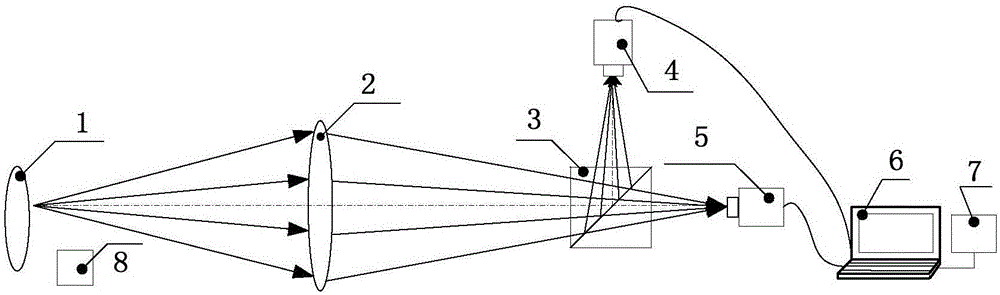
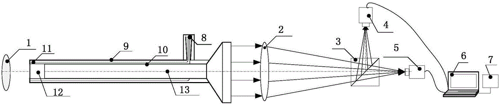
[0025] like figure 1 As shown, the positioning device for bulla rupture mainly includes: an imaging optical system 2, a dichroic beamsplitter prism 3, a first camera 4, a second camera 5, a control computer 6, a data processing system 7, and an active lighting source 8 fixed At the front end of the imaging optical system 2 (connecting structures such as a fixed frame are omitted in the figure), the observed alveolar tissue 1 is uniformly illuminated. The observed alveolar tissue 1 is illuminated by the active illumination light source 8, passes through the imaging optical system 2, and then passes through the dichroic beamsplitter prism 3, the visible light band is imaged on the target surface of the first camera, and the fluorescence band is imaged on the target surface of the second camera , the control computer is used to collect the image data of the first camera and the second camera, and the data processing system 7 is used to analyze and process the image data, display ...


PUM


Abstract

The invention provides a positioning device for a pulmonary bulla rupture site. According to the positioning device, a pulmonary alveoli rupture and air leakage site can be positioned quickly and accurately. The positioning device for the pulmonary bulla rupture site comprises an active lighting source, an imaging optical system, a dichroic light splitting prism, a first camera, a second camera, a control computer, a data handling system and a fluorescent material, wherein the first camera and the second camera are arranged on two-way light splitting light ways of the dichroic light splitting prism correspondingly, and the fluorescent material is used being matched with the active lighting source. An observed pulmonary alveoli tissue is under the irradiation of the active lighting source, through the imaging optical system, and through the dichroic light splitting prism, then a visible light wave band is imaged on the target face of the first camera, a fluorescence wave band is imaged on the target face of the second camera, the control computer collects image data of the first camera and the second camera, and the pulmonary alveoli rupture site is marked through the data handling system and displayed in real time on a display of the control computer. The system has a compact structure, a small volume and low cost, and a handheld portable type can be achieved.

Description

technical field [0001] The invention relates to an imaging detection device in the medical field, in particular to an imaging system structure for locating the location of alveolar rupture. Background technique [0002] Bullae secondary to pneumonia or lung abscess are more common in infants, single or multiple. Due to inflammatory lesions, the mucous membrane of the small bronchi is edematous, resulting in partial blockage of the lumen and a valve effect. Air can enter the alveoli and is not easy to be discharged. The pressure in the alveoli increases, and the alveolar septum gradually ruptures due to the increased pressure in the alveoli, forming a huge The air-sac-containing cavity in the lung, which is called bulla clinically, is one of the common emergencies in thoracic surgery. Small bullae themselves do not cause symptoms, and patients with simple bullae often have no symptoms. Some bullae can remain unchanged for many years, and some bullae can gradually increase. ...

Claims


Application Information

Patent Timeline
Patent Type & Authority Applications(China)
IPC IPC(8): A61B5/00A61B5/08
CPCA61B5/0059A61B5/0071A61B5/0082A61B5/08A61B2503/04A61B2503/06A61B2576/02
Inventor 张周锋于涛胡炳樑刘欢
Owner XI'AN INST OF OPTICS & FINE MECHANICS - CHINESE ACAD OF SCI
Why Eureka
Eureka Blog
Learn More