

Image processing method of hyperpolarized gas magnetic resonance
An image processing and magnetic resonance technology, which is applied in magnetic resonance measurement, measurement using nuclear magnetic resonance image system, and measurement of magnetic variables, etc., can solve the problems of difficulty and complexity in obtaining high-quality hyperpolarized gas magnetic resonance images, and achieve image The effect of detail information enhancement and measurement quality
- Summary
- Abstract
- Description
- Claims
- Application Information
AI Technical Summary
Problems solved by technology
Method used
Image

Examples
Embodiment 1
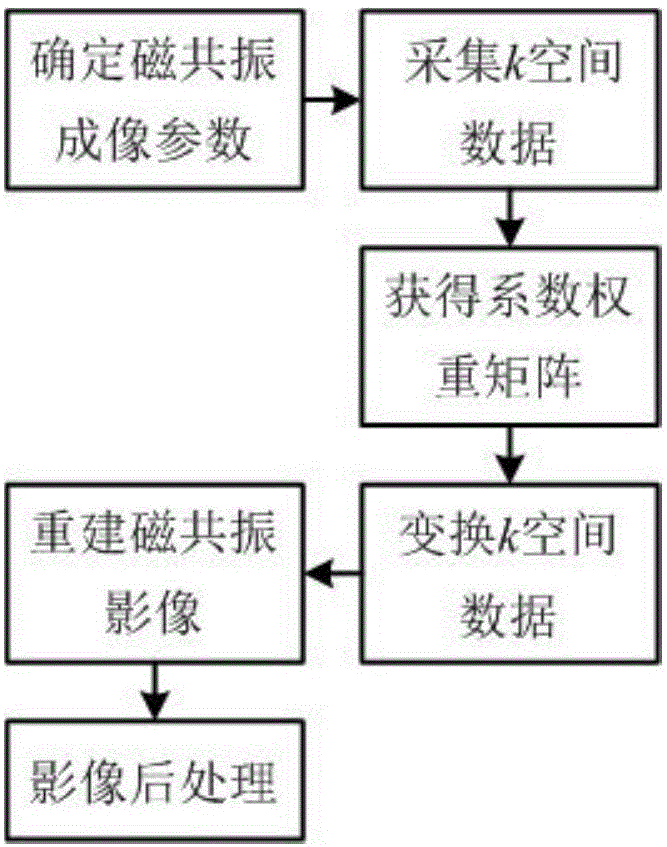
[0026] figure 1 It is a schematic block diagram of a hyperpolarized gas magnetic resonance image processing method of the present invention, mainly including - determination of hyperpolarized gas magnetic resonance imaging parameters (including echo time, repetition time, matrix size, number of layers, layer thickness, field of view, etc. ), k-space data acquisition (fixed-angle excitation / central encoding), coefficient weight matrix determination, k-space data transformation, magnetic resonance image reconstruction (inverse Fourier transform), and image post-processing (denoising), to obtain super Magnetic resonance imaging of polarized gases.
[0027] Specifically:
[0028] Step 1: Select the imaging sequence, set the echo time, and set the excitation angle to obtain the original hyperpolarized gas magnetic resonance k-space data.
[0029] Because the longitudinal magnetization vector of hyperpolarized gas magnetic resonance is non-reproducible, hyperpolarized gas magnetic...
Embodiment 2
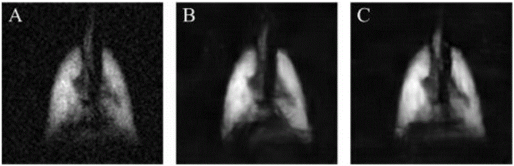
[0054] Such as image 3 As shown, steps 1-3 in this embodiment have the following differences from Embodiment 1: the imaging parameters of human lungs are: 1.5T magnetic resonance imager, the echo time is 2.7ms, the repetition time is 6.8ms, and the matrix size is 128×128, the number of layers is 7, the layer thickness is 20mm, and the field of view is 400×400mm 2 , the bandwidth is 25.6kHz, the total scan time is 6.1s, fixed-angle excitation (excitation angle is 9°), FLASH imaging sequence, center coding, the size of the coefficient weight matrix is 128×128, and the coefficient weight matrix is according to formula (7), The angle θ in the coefficient weight matrix is set to 6°. Others are the same as in Example 1.
[0055] image 3 A. image 3 B and image 3 C are the magnetic resonance images reconstructed according to the original hyperpolarized gas magnetic resonance k-space data of the fourth layer, the fifth layer and the sixth layer; image 3 D. image 3 E ...
Embodiment 3
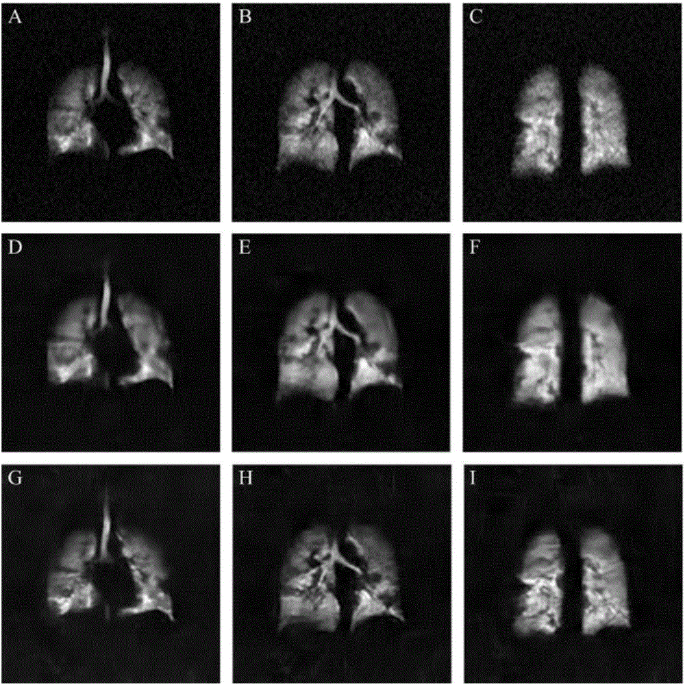
[0061] Such as Figure 4 As shown, steps 1-3 in this embodiment have the following differences from Embodiment 1: the imaging parameters of human lungs are: 1.5T magnetic resonance imager, the echo time is 2.7ms, the repetition time is 6.8ms, and the matrix size is 128×128, the number of layers is 8, the layer thickness is 20mm, and the field of view is 400×400mm 2 , the bandwidth is 25.6kHz, the total scan time is 6.97s, fixed-angle excitation (excitation angle is 9°), FLASH imaging sequence, center coding, coefficient weight matrix size is 128×128, coefficient weight matrix according to formula (7), The angle θ in the coefficient weight matrix is set to 6°. Others are the same as in Example 1.
[0062] Figure 4 A. Figure 4 B and Figure 4 C are the magnetic resonance images reconstructed according to the original hyperpolarized gas magnetic resonance k-space data of the fifth layer, the sixth layer and the seventh layer; Figure 4 D. Figure 4 E and Figure 4 F i...
PUM
 Abstract
Description
Claims
Application Information