Fast extraction of pelvic contours from serial CT images based on key frame markers

A CT image and key frame technology, applied in image analysis, image enhancement, image data processing, etc., can solve the problems of long operation time, sensitive initial contour, uneven bone density, etc. Good adaptability to differences and the effect of reducing image processing time

Active Publication Date: 2019-12-20
TIANJIN UNIV
View PDF3 Cites 0 Cited by
  • Summary
  • Abstract
  • Description
  • Claims
  • Application Information

AI Technical Summary

Problems solved by technology

[0003] In the bone CT image segmentation method, the most commonly used segmentation method is the segmentation method based on gray information, and the threshold method is a typical method, but the bone density is uneven, and the connection between the femoral head and the acetabulum is relatively narrow. , and weak margins caused by lesions, it is difficult to select an appropriate threshold during use
In addition, the classification and clustering methods in machine vision are also used in segmentation. This type of method has good robustness to noise, but the segmentation effect depends on the number and type of samples. Due to the large individual differences between patients , this type of segmentation algorithm has limitations
At present, a lot of research focuses on statistical shape model segmentation methods, such as snake model and GVF model. [1] (Gradient Vector ConvolutionField Snake Model), level set-based segmentation models, etc. The research focus of this type of segmentation method is on the automatic selection of marker points, the construction of models with fewer training sets, the improvement of the aforementioned models, and the combination with other methods, etc. On the one hand, a large amount of human processing is required to give prior information before the segmentation is realized. The final segmentation effect depends on the accuracy and completeness of the prior information. Due to the large individual differences between patients, to improve the segmentation effect, it is necessary to increase Prior information, so this kind of method has a lot of work in the early stage, but the effect is not guaranteed, so it is not suitable for direct use in hospitals
Due to the GVF model [2] It better solves two problems that the traditional snack model is difficult to solve: 1. It is very sensitive to the initial contour; 2. It cannot achieve the ideal effect when segmenting the concave part of the image
At present, a large number of studies have segmented images based on the GVF model, but the GVF model still has the shortcomings of being sensitive to the initial contour and taking a long time to calculate.

Method used


Image

  • Fast extraction of pelvic contours from serial CT images based on key frame markers
  • Fast extraction of pelvic contours from serial CT images based on key frame markers
  • Fast extraction of pelvic contours from serial CT images based on key frame markers

Examples


Embodiment Construction

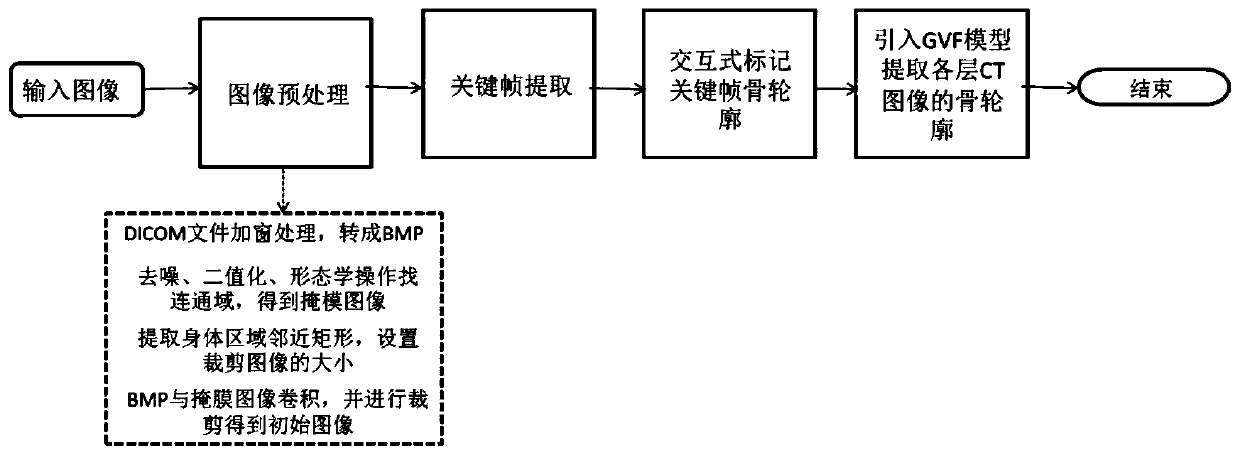
[0024] Considering that hospitals currently use manual marking of the pelvic region to formulate surgical plans and a patient has a large number of CT sequences, it takes about 15 minutes to manually mark a single CT image clinically, so manual segmentation of the pelvic region takes a long time and is stressful. Due to the small shooting distance in the sequential CT slices, there is little change in the morphological features of the bones between two adjacent frames, and there is a high similarity. Using this feature, the key frames are extracted from the CT sequence slices. After this step, it is necessary to segment The amount of data will be greatly reduced. By drawing the outline of the pelvic edge in the full CT sequence by the doctor marking the outline of the bone edge in a very small number of key frames, the segmentation of the entire pelvic region will be realized. This method not only reduces the processing time of the doctor, but also It can adapt to individual di...


PUM


Abstract

The invention relates to a sequence CT image pelvis contour extraction method based on keyframe tagging. The method comprises the steps of conducting preprocessing on CT images; obtaining a mask of an area-of-interest; conducting convolution on the mask and an initial image to obtain intermediate images; ranking the intermediate images according to a spatial sequence, selecting a pixel difference value of two adjacent frames of CT images as a feature distance, regarding two adjacent frames of which the feature distance is smaller than a given threshold value as similar frames, and conducting preliminary screening on a CT sequence to obtain a candidate keyframe sequence; conducting fine screening on the candidate keyframe sequence; tagging keyframe bone contour in an interactive mode; adopting a GVF model to extract the bone contour of the CT images of all layers.

Description

technical field [0001] The invention relates to the field of medical image segmentation, in particular to a method for segmenting bone regions such as the great pelvis, sacrum, hip bone, and acetabulum in CT images of the pelvis. Background technique [0002] Medical image segmentation is the basis of medical image analysis and processing. The accuracy of medical image segmentation will directly affect the doctor's judgment of the disease and the choice of surgical plan. Due to the high resolution of CT images, which can clearly display the characteristics of anatomical structures and diseased tissue areas, CT has been widely used in the diagnosis of various diseases. Pelvic fractures are one of the factors causing morbidity and mortality. For displaced fractures, accurately and quickly determining the extent of the fracture, the degree of comminution, and the degree of soft tissue damage can provide a reference for the selection of treatment methods and the prognosis. Seco...

Claims


Application Information

Patent Timeline
Patent Type & Authority Patents(China)
IPC IPC(8): G06T7/12G06T7/13G06T7/181G06T5/00
CPCG06T5/002G06T5/006G06T2207/10081G06T2207/30008
Inventor 余辉王海均张力新孙敬来曹玉珍于旭耀时尧
Owner TIANJIN UNIV
Why Eureka
Try Eureka