Method for separating exosomes
A separation method and technology of exosomes, applied in the biological field, can solve problems such as quality reduction, vesicle damage, and time-consuming process, and achieve high yield, reduced centrifugation time, and good results
- Summary
- Abstract
- Description
- Claims
- Application Information
AI Technical Summary
Problems solved by technology
Method used
Image
Examples
Embodiment 1
[0059] The construction of embodiment 1 plasmid
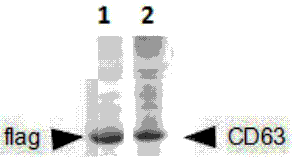
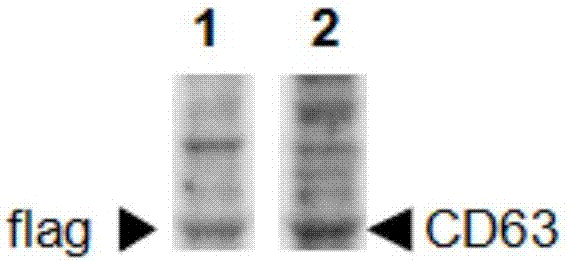
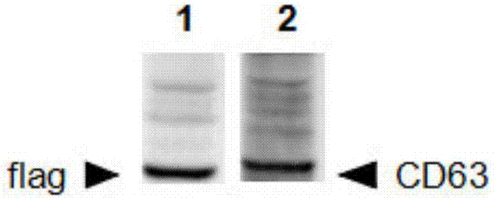
[0060] The plasmid pCDNA3.1 was digested with Nhe I endonuclease and restriction endonuclease Hind III, and a band with a vector size of 5.4kb was recovered; the primer CD63-F / R amplified the synthesized CD63 template to obtain a CD63 fragment; because 3flag-F / R are templates for each other, and the 3flag fragment is obtained by PCR amplification. The vector, CD63 fragment and 3flag fragment were added to the HB-infusion seamless cloning kit premix at a molar ratio of 1:3:3, and ligated to obtain the target plasmid pCD63-3*Flag.
[0061] Specific components of the medium in Table 1
[0062]
Embodiment 2
[0063] Example 2 Isolation of exosomes in 293 cells
[0064] 1) Cultivate 293 cells, and when the cell density reaches 70-80%, transfer pCD63-3*Flag into the cells by PEI method, and after culturing for 24 hours, use G418 to screen out the positive clone 293-CD63-3*Flag cells; 293-CD63-3*Flag cells were planted in a 100mm culture dish and cultured for 1 day using the aforementioned cell culture medium;
[0065] 2) Wash the cells with serum-free a-MEM, 5 times, 5ml each time;
[0066] 3) Culture with 10ml 10% a-MEM without exosomes FBS (fetal bovine serum) for 3 days, collect the supernatant, centrifuge at 1000g for 5min, and then centrifuge at 12000g for 30min at 4°C to remove broken cell debris and apoptotic bodies;
[0067] 4) After centrifugation, mix the supernatant with PBS at a volume ratio of 1:1;
[0068] 5) Add flag antibody-coated beads (magnetic beads) to the medium solution containing exosomes, incubate at 4°C for 15 minutes, and the magnetic beads bind to exosom...
Embodiment 3
[0074] Example 3 Isolation of exosomes in Hela cells
[0075] 1) Cultivate Hela cells, and when the cell density reaches 70-80%, transfer pCD63-3*Flag into the cells by PEI method, and after culturing for 24 hours, use G418 to screen out the positive clone Hela-CD63-3*Flag cells; 2 *10^6 Hela-CD63-3*Flag cells were planted in a 100mm culture dish and cultured for 1 day using the aforementioned cell culture medium;
[0076] 2) Wash the cells with serum-free a-MEM, 5 times, 5ml each time;
[0077] 3) Culture with 10ml 10% a-MEM without exosome FBS for 3 days, collect the supernatant, centrifuge at 1000g for 5min, then centrifuge at 12000g at 4 degrees for 30min to remove broken cell debris and apoptotic bodies;
[0078] 4) After centrifugation, mix the supernatant with PBS at a ratio of 1:1;
[0079] 5) Add flag antibody-coated beads (magnetic beads) to the medium solution containing exosomes, incubate at 2°C for 20 minutes, and the magnetic beads bind to exosomes with corresp...
PUM
 Abstract
Description
Claims
Application Information
 - R&D
- Intellectual Property
- Life Sciences
- Materials
- Tech Scout
- Unparalleled Data Quality
- Higher Quality Content
- 60% Fewer Hallucinations
Browse by: Latest US Patents, China's latest patents, Technical Efficacy Thesaurus, Application Domain, Technology Topic, Popular Technical Reports.
© 2025 PatSnap. All rights reserved.Legal|Privacy policy|Modern Slavery Act Transparency Statement|Sitemap|About US| Contact US: help@patsnap.com


