OCT endoscopic imaging probe and manufacturing method thereof

A technology of imaging probe and manufacturing method, which is applied in the field of probe, can solve problems such as decomposition, attenuation, and influence of infrared spot quality, and achieve the effect of optimizing focus

Active Publication Date: 2017-08-08
INNERMEDICAL CO LTD
View PDF5 Cites 6 Cited by
  • Summary
  • Abstract
  • Description
  • Claims
  • Application Information

AI Technical Summary

Problems solved by technology

But this structure has three disadvantages: 1. The quality of the infrared spot coming out of the probe fiber is affected by the sheath at the end of the probe; 2. The signal light returned from the biological tissue is severely attenuated when it passes through the sheath at the end of the probe; 3. The optical fiber probe uses glue Attached to a torque coil, the adhesive softens and even breaks down when the probe is autoclaved

Method used


Image

  • OCT endoscopic imaging probe and manufacturing method thereof
  • OCT endoscopic imaging probe and manufacturing method thereof

Examples


Embodiment Construction

[0029] The present invention will be further described below in conjunction with the accompanying drawings and specific embodiments.

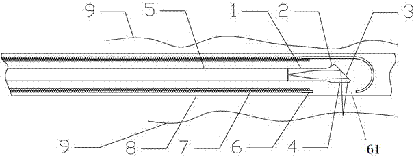
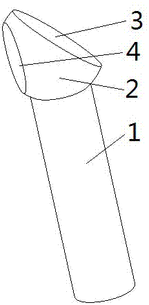
[0030] Such as figure 1 As shown, an OCT endoscopic imaging probe includes a self-focusing optical fiber 1, the light-emitting end of the self-focusing optical fiber 1 is connected with a ball lens 2, and the ball lens 2 is provided with a reflective surface 3 and is connected to the reflective surface 3. Corresponding to the light exit window 4, the light emitted by the self-focusing optical fiber 1 is emitted through the light exit window 4 under the reflection of the reflective surface 3. The size of the reflective surface 3 is preferably capable of completely reflecting the speed of light, so The size of the light output window 4 is to meet the requirements of the light aperture, the reflective surface 3 and the light output window 4 are both plane, and the OCT endoscopic imaging probe can perform imaging work in the blood vessel wall 9 . ...


PUM


Abstract

The invention provides an OCT endoscopic imaging probe. The OCT endoscopic imaging probe comprises a self-focusing optical fiber, wherein a spherical lens is connected to the light-outgoing end of the self-focusing optical fiber, a reflecting surface and a light-outgoing window corresponding to the reflecting surface are arranged on the spherical lens, light emitted from the self-focusing optical fiber is projected through the light-outgoing window under the reflecting effect of the reflecting surface, and the reflecting surface and the light-outgoing window are planes. The invention further provides a manufacturing method of the OCT endoscopic imaging probe. The OCT endoscopic imaging probe has the beneficial effects that the focusing of light can be optimized, the signal attenuation can be avoided, and the firmness of the probe is enhanced.

Description

technical field [0001] The invention relates to a probe, in particular to an OCT endoscopic imaging probe and a manufacturing method thereof. Background technique [0002] Optical coherence tomography (Optical Coherence Tomography, OCT) uses near-infrared light and the principle of optical interference to detect the back reflection or several scattering signals of incident near-infrared light at different depths of biological tissues. Through scanning, it can be obtained Two-dimensional or three-dimensional structural images of biological tissues. In recent years, although it has been found that it has the advantages of non-destructive and high resolution in vascular imaging and intracavitary tissue (stomach) examination, it is difficult to achieve large-scale application due to problems such as complex manufacturing process and poor imaging effect. [0003] At present, most OCT probes on the market adopt the following two implementation methods: [0004] One is to bond gl...

Claims


Application Information

Patent Timeline
Patent Type & Authority Applications(China)
IPC IPC(8): A61B5/00A61B1/07A61B1/05
CPCA61B1/00131A61B1/05A61B1/0661A61B1/07A61B5/0066A61B5/0086A61B5/6847A61B5/687A61B5/6871A61B5/6876
Inventor 白晓淞
Owner INNERMEDICAL CO LTD
Why Eureka
Try Eureka