Attaching-type heart function monitoring and/or intervening system
A heart function and heart technology, applied in the field of medical devices, can solve the problems of inability to perform quantitative, timing, real-time, anytime, and timely artificial regulation, clinical efficacy of heart failure treatment devices, limited application potential, and difficulty in adjustment and control.
- Summary
- Abstract
- Description
- Claims
- Application Information
AI Technical Summary
Problems solved by technology
Method used
Image
Examples
Embodiment 1
[0062] Example 1 Preparation of Heart Mesh Cover
[0063] (1) Carry out computer-aided design (CAD) modeling using methods routinely used in this field. These designs can be derived from digital image reconstructions of the patient's heart. For example, image data can be obtained through non-invasive scanning of the human body (such as MRI or CT) or fine-layered three-dimensional reconstruction;
[0064] (2) Use liquid silicone, latex, conductive hydrogel, silicone glue, rubber or polymer plastic materials, and use 3D printing technology to print the heart mesh cover;
[0065] or,
[0066] ① Use blue wax, green wax, red wax, black wax, white wax and other materials to make the solid structure of the device with 3D printing equipment;
[0067] ② Soak the blue wax solid structure in liquid silica gel or latex or conductive hydrogel or silicone glue or rubber or polymer plastic material for 1 second to 24 hours;
[0068] ③Take out the above soaked structure, cover it with a c...
Embodiment 2
[0072] Example 2 Attached heart function monitoring system
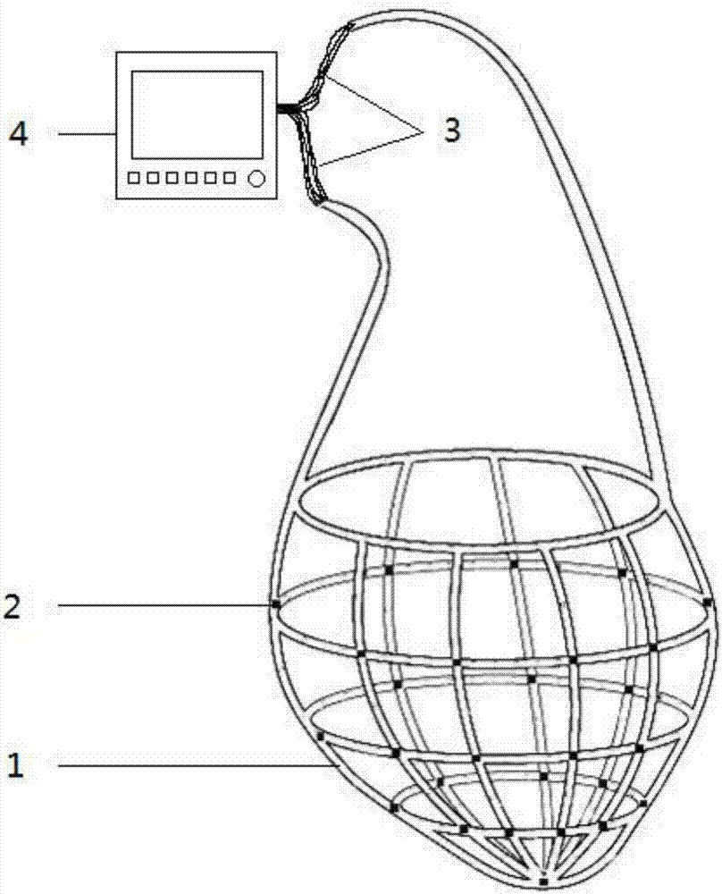
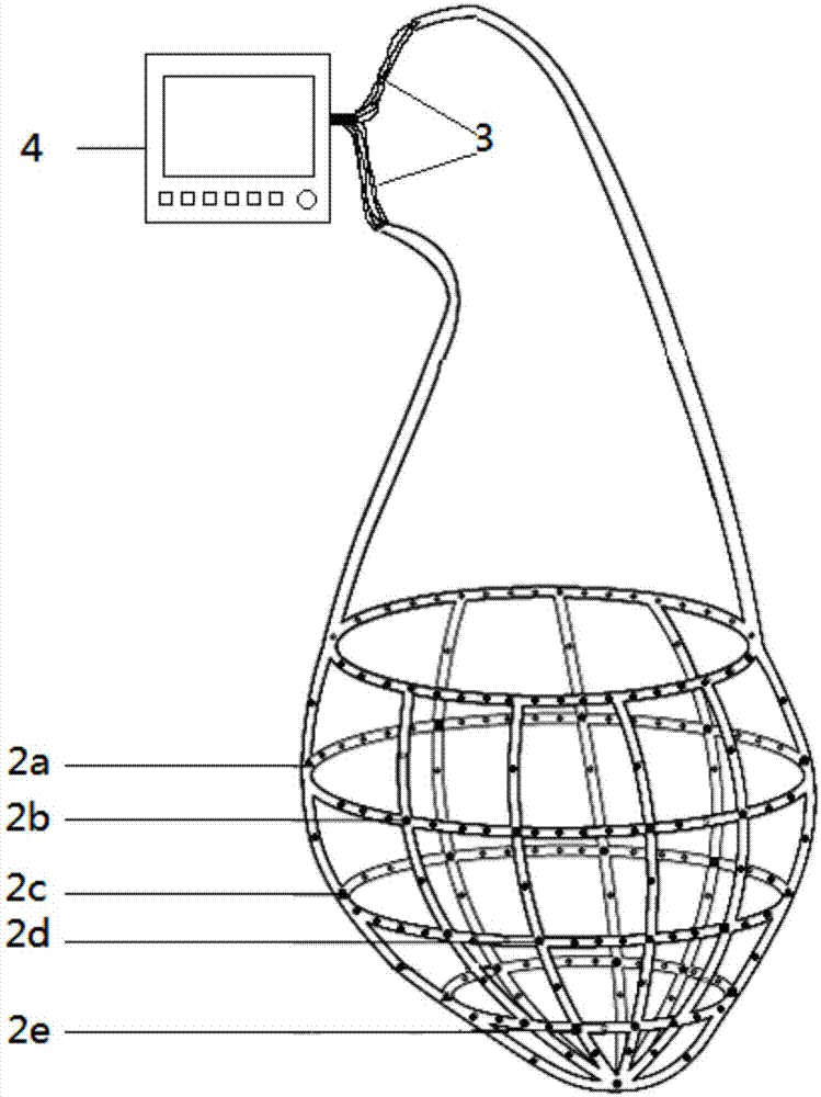
[0073] An attached heart function monitoring and / or intervention system, a heart support device and a heart function monitoring device, the heart support device is selected from a heart mesh sleeve, and the structure is as follows figure 1 As shown, 1-cardiac mesh, 2-physiological and biochemical sensors, 3-wire, 4-heart function monitoring device. The cardiac support device is coated on the outer surface of the ventricle and / or atrium or the support is attached to the inner surface of the cardiac cavity, the cardiac function monitoring device is connected with physiological and biochemical sensors, and the physiological and biochemical sensors are attached to the inner and / or outer surface of the cardiac support device , or embedded in the cardiac support device, or filled inside the cardiac support device.
Embodiment 3
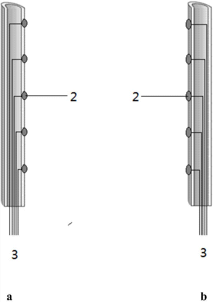
[0075] The basic structure is the same as that of Example 2. The net cover is composed of hollow tubes. All the hollow tubes are completely connected or form several independent areas. The inside of the area is interconnected, but the areas are not connected. The physiological and biochemical sensor paste Attached to the inside or outside of the net, the wires of the physiological and biochemical sensors pass through the hollow tube of the net and are connected to the heart function monitoring device from the end of the net. structured as figure 2 As shown, when the mesh is attached to the outer surface of the ventricle and / or atrium, the physiological and biochemical sensors are attached to the inside of the mesh ( figure 2 a), when the mesh sleeve is attached to the inner surface of the heart cavity, the physiological and biochemical sensors are attached to the outside of the mesh sleeve ( figure 2 b).
PUM
 Abstract
Description
Claims
Application Information
 - R&D
- Intellectual Property
- Life Sciences
- Materials
- Tech Scout
- Unparalleled Data Quality
- Higher Quality Content
- 60% Fewer Hallucinations
Browse by: Latest US Patents, China's latest patents, Technical Efficacy Thesaurus, Application Domain, Technology Topic, Popular Technical Reports.
© 2025 PatSnap. All rights reserved.Legal|Privacy policy|Modern Slavery Act Transparency Statement|Sitemap|About US| Contact US: help@patsnap.com


