
Local ablation method and system for liver cancer

A local, liver cancer technology, applied in surgical navigation systems, image data processing, heating surgical instruments, etc., can solve problems such as inconsistencies, and achieve the effect of improving positioning accuracy, objective and convenient

Inactive Publication Date: 2017-08-18
纪建松
View PDF7 Cites 45 Cited by
  • Summary
  • Abstract
  • Description
  • Claims
  • Application Information

AI Technical Summary

Problems solved by technology

However, sometimes the results of ultrasound, CT, and MRI examinations are inconsistent, and clinicians are required to be familiar with various imaging manifestations after tumor ablation, so as to accurately evaluate the treatment effect and detect new and recurrent lesions

Method used


Image

  • Local ablation method and system for liver cancer
  • Local ablation method and system for liver cancer
  • Local ablation method and system for liver cancer

Examples


Embodiment 1

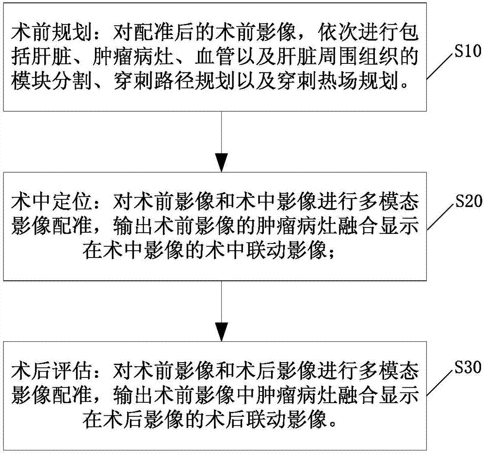
[0063] Such as figure 1 As shown, the present invention provides a method for local ablation of liver cancer, which comprises the steps of:

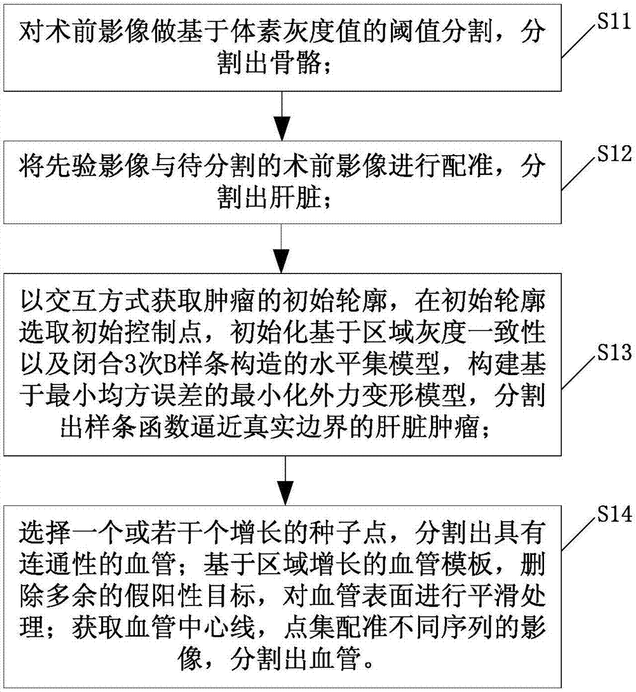
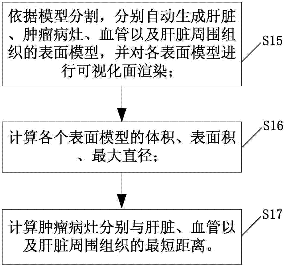
[0064] S10, preoperative planning: for the pre-registered preoperative images, sequentially perform module segmentation including liver, tumor lesions, blood vessels, and tissues around the liver, puncture path planning, and puncture thermal field planning.

[0065] S20, intraoperative positioning: multimodal image registration is performed on the preoperative image and the intraoperative image, and the tumor lesion output from the preoperative image is fused and displayed on the intraoperative linkage image of the intraoperative image;

[0066] S30, postoperative evaluation: multimodal image registration is performed on the preoperative image and postoperative image, and the fusion of tumor lesions in the output preoperative image is displayed on the postoperative image.

[0067] In the above step S10, the preoperative planning is main...

Embodiment 2

[0076] On the basis of Example 1, this embodiment of the present invention provides a local liver cancer ablation system, such as Figure 8(a)-Figure 8(b) shown, which includes:

[0077] Preoperative planning unit 10: for the registered preoperative images, it sequentially performs the module segmentation module including the liver, tumor lesions, blood vessels and tissues around the liver, the puncture path planning module for planning the puncture path, and the puncture thermal field path planning module. A puncture thermal field planning module for planning;

[0078] Intraoperative positioning unit 20, which performs multimodal image registration on preoperative images and intraoperative images, and outputs tumor lesions in preoperative images that are fused and displayed in intraoperative linkage images of intraoperative images;

[0079] A postoperative evaluation unit 30, which performs multimodal image registration on the preoperative image and the postoperative image, ...


PUM


Abstract

The invention discloses a local ablation method for liver cancer. The local ablation method comprises the following steps: carrying out modular partition including the liver, the tumor focus, the blood vessel and the tissue around the liver in sequence on the preoperative image after registration, and carrying out puncture path planning and puncture thermal field planning; carrying out multi-modal image registration on the preoperative image and an intra-operative image, outputting the tumor focus of the preoperative image, and displaying the tumor focus on the intra-operative image in a fusion manner; and carrying out multi-modal image registration on the preoperative image and a postoperative image, outputting the tumor focus in the preoperative image, and displaying the tumor focus in the preoperative image on the postoperative image in a fusion manner, wherein the preoperative image and the postoperative image respectively comprise the CT image and / or MR image of the time sequence, and the intra-operative image comprises the CT image. According to the method, the image registration step is added in preoperative planning, the intra-operative locating and postoperative assessment, thus the scientific planning for the tumor thermal ablation before the operation, the visual and effective information display in treatment during the operation, and the scientific assessment for the curative effects and the treatment experience feedback after the operation are realized, so that the preoperative planning is guided.

Description

technical field [0001] The present invention relates to the technical field of surgical local ablation, and more specifically, the present invention relates to a method and system for local ablation of liver cancer. Background technique [0002] my country is a country with a high incidence of liver cancer, accounting for more than half of the world's annual incidence and death, which seriously threatens the health and lives of our people. Surgical resection is the treatment of choice for liver cancer. However, surgical resection cannot be performed for patients with massive HCC, large number of tumors, vascular invasion such as portal vein and hepatic vein, extrahepatic metastasis, and poor liver reserve. With the advancement of medical imaging and interventional therapy, minimally invasive treatment of local tumors has developed rapidly, including percutaneous ethanol and acetic acid injection, microwave ablation, radiofrequency ablation and argon-helium knife ablation. ...

Claims


Application Information

Patent Timeline
Patent Type & Authority Applications(China)
IPC IPC(8): A61B18/12A61B34/10A61B34/20G06T5/00G06T7/00G06T15/00
CPCG06T15/00A61B18/12G06T2207/10088G06T2207/10081G06T2207/30096G06T2207/30056G06T5/70
Inventor 纪建松涂建飞周志勇徐民赵中伟
Owner 纪建松
Learn More