Fundus camera

A camera and fundus image technology, which is applied in the field of medical imaging, can solve problems such as increased information input, medical staff struggling to cope with information input work, and inability to concentrate on fundus photography and fundus image analysis to achieve the effect of reducing workload

Inactive Publication Date: 2017-08-25
SUZHOU MICROCLEAR MEDICAL INSTR
View PDF9 Cites 1 Cited by
  • Summary
  • Abstract
  • Description
  • Claims
  • Application Information

AI Technical Summary

Problems solved by technology

When the flow of patients is large, the corresponding amount of information input will increase year-on-year, which makes the medical staff tired of coping with the information input work, and cannot concentrate on fundus photography and fundus image analysis.

Method used


Image

  • Fundus camera
  • Fundus camera

Examples


Embodiment Construction

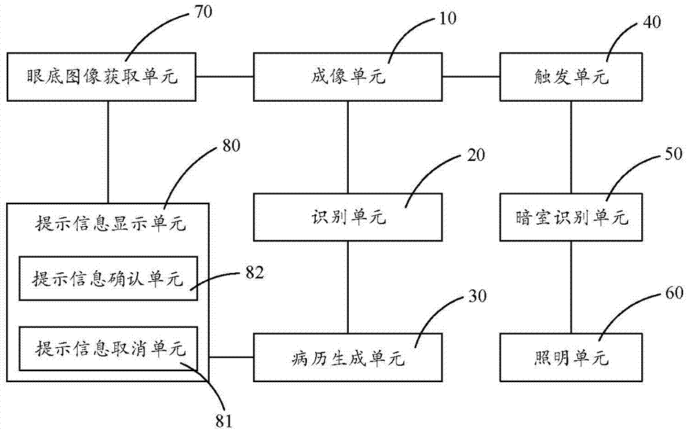
[0020] see figure 1 As shown, the fundus camera of the present invention includes an imaging unit 10 , an identification unit 20 and a medical record generation unit 30 . The imaging unit 10 is used to take pictures of the patient's ID card and fundus to obtain image information such as ID images and fundus images. The recognition unit 20 is used to recognize the identification information recorded on the certificate image. The identification information includes name, ID number, photo and so on. The medical record generation unit 30 generates an electronic medical record according to the identification information, so that each fundus image can be associated with an electronic medical record, which is convenient for medical personnel to identify, effectively reduces the workload of medical personnel, and allows medical personnel to concentrate on In fundus photography and fundus image analysis work.

[0021] Preferably, the fundus camera also has a trigger unit 40 . When ...


PUM


Abstract

The invention discloses a fundus camera, comprising: an imaging unit, which is used for taking pictures of a patient's ID card and the fundus to obtain image information such as the ID image and the fundus image; and an identification unit, which is used for identifying all identification information recorded on the certificate image; a medical record generating unit, the medical record generating unit generates an electronic medical record according to the identification information. Compared with the prior art, the fundus camera of the present invention can easily associate the fundus image with the patient information without manual input, thereby greatly reducing the workload of the medical staff, so that the medical staff can concentrate on the fundus photography and the fundus image. analyze.

Description

technical field [0001] The invention relates to the field of medical imaging, in particular to a fundus camera. Background technique [0002] The fundus camera belongs to the field of medical imaging and is used to obtain retinal images of the human eye so that medical personnel can check fundus diseases or assist medical personnel to judge the condition of other organs. Since the blood vessels of the fundus are the only blood vessels that can be directly observed through the body surface, medical personnel can check whether there are lesions in the optic nerve, retina, choroid and refractive medium of the fundus through the fundus camera, and can also assist in the treatment of other system diseases through the fundus camera Diagnosis and disease judgment, such as detecting cerebral infarction, cerebral hemorrhage, cerebral arteriosclerosis, brain tumor, diabetes, kidney disease, hypertension, retinopathy of prematurity, glaucoma, age-related macular degeneration, etc. by s...

Claims


Application Information

Patent Timeline
Patent Type & Authority Applications(China)
IPC IPC(8): A61B3/12A61B3/14
CPCA61B3/12A61B3/0016A61B3/14
Inventor 晏虎周亮亮李超宏
Owner SUZHOU MICROCLEAR MEDICAL INSTR
Why Eureka
Try Eureka