Breast tissue segmentation method

A technology of breast tissue and lung tissue, which is applied in the field of medical image processing, can solve the problems of initial position sensitivity, segmentation result deviation, and susceptibility to noise interference, and achieve high positioning accuracy, accurate segmentation, and high accuracy.

Active Publication Date: 2017-08-29
SHANGHAI UNITED IMAGING HEALTHCARE
View PDF5 Cites 19 Cited by
  • Summary
  • Abstract
  • Description
  • Claims
  • Application Information

AI Technical Summary

Problems solved by technology

Among them, the classic threshold method identifies and extracts the target from the background by finding the local optimal value or the global optimal value. There is a certain deviation in the segmentation results obtained by the method; the watershed transform can generate a single-pixel closed contour when segmenting the image by segmenting the gradient image, but there is an over-segmentation phenomenon; the Snake model can achieve the extraction of the target contour, but it is sensitive to the initial position , easy to fall into local extremum

Method used


Image

  • Breast tissue segmentation method
  • Breast tissue segmentation method
  • Breast tissue segmentation method

Examples


Embodiment Construction

[0046] To make the above objects, features and advantages of the present invention more obvious and comprehensible, specific implementations of the present invention will be described in detail below in conjunction with the accompanying drawings and embodiments.

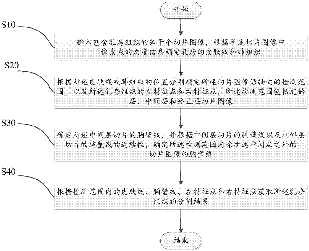
[0047] A method for segmenting breast tissue of the present invention is as follows: figure 1 shown, including the following steps:

[0048] S10. Input several or several slice images (or slice images) including breast tissue, and determine breast skin line and lung tissue according to grayscale information of pixels in the slice images. The imaging method of breast tissue slice images can be magnetic resonance equipment (MR) imaging, computerized tomography (CT), X-ray machine (especially breast machine) imaging, etc. Specifically, in this embodiment, breast tissue CT Taking a slice image as an example, the CT slice image may also be called a three-dimensional sequence image, which includes several slices (images)....


PUM


Abstract

The invention discloses a breast tissue segmentation method comprising the steps that multiple section images including breast tissues are inputted, and the skin line of the breasts and the lung tissues are determined according to grayscale information of the pixel points in the section images; the detection range of the section images along an axial direction and the left and right feature points of the breast tissues are determined according to the position of the skin line or the lung tissues, wherein the detection range includes the initial layer, intermediate layer and stop layer section times; the chest wall line of the intermediate layer section is determined, and the chest wall line of the section images apart from the intermediate layer section in the detection range is determined according to the continuity of the chest wall line of the intermediate layer section and the chest wall line of the adjacent layer sections; and the segmentation result of the breast tissues is acquired according to the skin line, the chest wall line, the left feature point and the right feature point in the detection range. According to the technical scheme, the position of the chest wall line of the breast tissues can be accurately and rapidly determined so as to realize automatic segmentation of the breast tissues.

Description

【Technical field】 [0001] The invention relates to the field of medical image processing, in particular to a breast tissue segmentation method. 【Background technique】 [0002] Breast disease is a common disease that threatens women's health, and its morbidity and mortality rank first among women's diseases. With the rapid development of modern medical diagnosis, the application of computer technology-assisted detection technology in the diagnosis of early breast diseases has been continuously improved. The relevant imaging examinations mainly include ultrasound, X-ray, magnetic resonance (MR), positron Emission scanning (PET) and computerized tomography (CT), etc., can obtain intuitive views and various quantitative and qualitative indicators of lesions by positioning and segmenting images after imaging examinations, which can be used for further diagnosis and treatment, Provide reference for preoperative prediction and intraoperative detection. At the same time, in the rad...

Claims


Application Information

Patent Timeline
Patent Type & Authority Applications(China)
IPC IPC(8): G06T7/10
CPCG06T2207/30068
Inventor 姜娈李鹏程李强
Owner SHANGHAI UNITED IMAGING HEALTHCARE
Why Eureka
Try Eureka