Method and device for displaying marker point inside three-dimensional medical model and medical equipment

A marking point and medical technology, which is applied in the field of medical display, can solve the problems of being unable to distinguish the real position of the marking point, unable to distinguish whether the marking point is inside or outside the three-dimensional medical model, and unable to effectively compare and view it, so as to achieve the effect of improving accuracy

Active Publication Date: 2017-10-10
QINGDAO HISENSE MEDICAL EQUIP
View PDF7 Cites 6 Cited by
  • Summary
  • Abstract
  • Description
  • Claims
  • Application Information

AI Technical Summary

Problems solved by technology

[0005] Due to the transparency of the 3D medical model, the visual positions of the marker points inside and on the surface of the 3D medical model are the same in the display results, and it is impossible to d...

Method used


Image

  • Method and device for displaying marker point inside three-dimensional medical model and medical equipment
  • Method and device for displaying marker point inside three-dimensional medical model and medical equipment
  • Method and device for displaying marker point inside three-dimensional medical model and medical equipment

Examples


Embodiment 1

[0092] refer to figure 2 As shown, a method for displaying internal marker points of a three-dimensional medical model provided by an embodiment of the present invention includes the following steps:

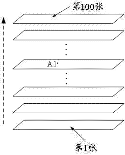
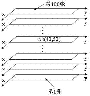
[0093] Step 201: Configure a marker point inside the 3D medical model as a virtual point light source, wherein the marker point represents a lesion point in a 2D medical image.

[0094] The virtual point light source is a virtual point light source capable of emitting light. For example, corresponding optical parameters can be configured through graphics software OpenGL (English: Open Graphics Library) to realize the simulation of point light source emitting light. The virtual point light source configured by OpenGL can emit light according to preset parameters to achieve the luminous effect of simulating a real point light source.

[0095] Specifically, if the lesion position marked by the doctor in the 2D medical image corresponds to the marked point in the 3D medical model ...

Embodiment 2

[0110] refer to Figure 3A As shown, a method for displaying internal marker points of a three-dimensional medical model provided by an embodiment of the present invention includes the following steps:

[0111] Step 301: According to the preset luminous intensity and attenuation coefficient, configure the mark point inside the three-dimensional medical model as a virtual point light source, wherein the mark point represents the lesion point in the two-dimensional medical image.

[0112] Specifically, if the lesion position marked by the doctor in the 2D medical image corresponds to the marked point in the 3D medical model inside the 3D medical model, configure the marked point inside the 3D medical model as a virtual point light source.

[0113] As an example, corresponding optical parameters can be configured through graphics software OpenGL (English: Open Graphics Library) to simulate light from a point light source. The virtual point light source configured by OpenGL can e...

Embodiment 4

[0195] refer to Figure 5 As shown, a method for displaying internal marker points of a three-dimensional medical model provided by an embodiment of the present invention includes the following steps:

[0196] Step 501: According to the preset luminous intensity, cutoff angle and attenuation coefficient, configure the mark point inside the 3D medical model as a virtual point light source, wherein the mark point represents the lesion point in the 2D medical image.

[0197] It should be noted that, for the execution process of step 501, reference may be made to the previous steps 201, 301, and 401, and the embodiment of the present invention will not be repeated here.

[0198] Step 502: configure the inner surface of the three-dimensional medical model so that the reflectivity is greater than the refractive index, wherein the three-dimensional medical model is a hollow structure.

[0199] It should be noted that the execution process of step 502 is the same as the execution pro...


PUM

PropertyMeasurementUnit
Light intensityaaaaaaaaaa

Abstract

The invention provides a method and device for displaying a marker point inside a three-dimensional medical model and medical equipment, and belongs to the technical field of medical display. The method comprises the steps that the marker point inside the three-dimensional medical model is configured to be a virtual point light source; a local fluoroscopy window is configured on the surface of the three-dimensional medical model; a light spot formed by the virtual point light source in the fluoroscopy window is utilized to represent the depth of the marker point inside the three-dimensional medical model, the depth of the marker point inside the three-dimensional medical model can be visually reflected through the light spot, and a doctor can further visually distinguish the marker point located inside or on the surface of the three-dimensional medical model so that depth information of the marker point inside the three-dimensional medical model can be obtained when the doctor conducts comparative viewing, which is beneficial to improving the accuracy of computer aided medical diagnosis i.

Description

technical field [0001] Embodiments of the present invention relate to the technical field of medical display, and in particular to a method, device and medical equipment for displaying internal marker points of a three-dimensional medical model. Background technique [0002] With the development and application of medical imaging technologies such as computed tomography (CT), magnetic resonance imaging (MRI), and ultrasound (US), traditional two-dimensional images only express the anatomical information of a certain section, while three-dimensional visualization technology Three-dimensional and spatial information constructs the relationship between the size and shape of human organs, soft tissues and lesions, and the location of surrounding tissues, which can assist doctors in diagnosis and treatment more vividly and three-dimensionally, and improve the accuracy and efficiency of diagnosis and treatment planning. Therefore, 3D visualization technology is becoming more and mo...

Claims


Application Information

Patent Timeline
IPC IPC(8): A61B90/00
CPCA61B90/39
Inventor 韩月乔田广野
Owner QINGDAO HISENSE MEDICAL EQUIP
Why Eureka
Try Eureka