Bladder volume measurement and calculation method under abdominal two-dimensional ultrasound

A technology of volume measurement and calculation method, which is applied in the field of bladder volume calculation, can solve the problems of high hardware equipment requirements, complex decomposition, high inspection cost, etc., and achieve the effects of ensuring practicability and accuracy, saving operation time, and simple measurement process

Active Publication Date: 2017-11-03
CHINA THREE GORGES UNIV
View PDF5 Cites 4 Cited by
  • Summary
  • Abstract
  • Description
  • Claims
  • Application Information

AI Technical Summary

Problems solved by technology

Although this technology converts two-dimensional space images into three-dimensional space data and enhances computing power, its shortcomings are also obvious. It requires high hardware equipment and requires three-dimensional volume probes and related software support, and usually has three-dimensional volume functions. The ultrasonic instrument is large in size, not easy to carry and move, and the inspection cost is high; the operator must be proficient in the relevant operating techniques of three-dimensional volume imaging; the entire operation and calculation cycle is time-consuming and time-sensitive
[0006] At present, there are portable ultrasonic bladder volume measuring instruments on the market. The principle is that after two-dimensional or three-dimensional imaging, the software automatically calculates the boundary of the inner wall of the bladder to obtain the corresponding image perimeter and area data, and then inserts the data into the existing mathematical formula. , and finally the bladder capacity is obtained by combining the data of each area. This method has high requirements for hardware equipment, and the corresponding ultrasound equipment needs to be purchased separately, and there are related conditions in the process of use, such as those with surgical scars on the abdomen and those who are too obese. etc., and it is useless for objects beyond its scanning area
[0007] To find the volume of an irregular object from the perspective of calculus, it is necessary to subdivide the object into n small units for calculation. This method not only requires the relevant mathematical operation ability and foundation, but also has very high requirements for hardware equipment. Sampling the bladder with fine three-dimensional heavy-dimensional data, the decomposition is complicated, the calculation time is too long, the timeliness is extremely poor, and the clinical application is unrealistic

Method used


Image

  • Bladder volume measurement and calculation method under abdominal two-dimensional ultrasound
  • Bladder volume measurement and calculation method under abdominal two-dimensional ultrasound
  • Bladder volume measurement and calculation method under abdominal two-dimensional ultrasound

Examples


Embodiment 1

[0048] Such as Figure 1~7 Shown, a kind of bladder volume measurement and calculation method under transabdominal two-dimensional ultrasound, it comprises the following steps:

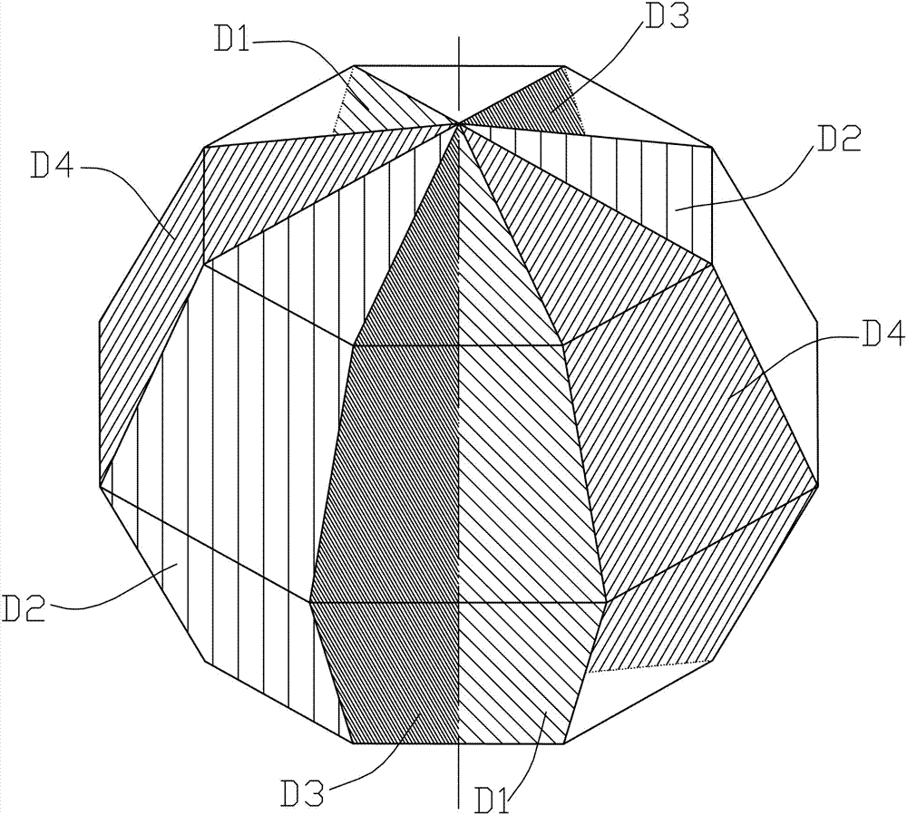
[0049] Step1: Scan the bladder through the abdomen, obtain a two-dimensional image of the bladder by ultrasound, fix the display depth of the acoustic window, try to distinguish the boundary of the bladder as clearly as possible, pressurize the probe appropriately, and stabilize the force to display the bladder as much as possible without affecting the general shape of the bladder The largest section of the median sagittal plane, that is, the median sagittal plane D1;

[0050] Step2: Rotate the probe counterclockwise by 90°, repeat the steps in Step1, and obtain the largest cross-section of the bladder, that is, cross-section D2;

[0051] Step3: Rotate the probe 45° clockwise on the basis of D1, and repeat the steps in Step1 to obtain the largest section of the left oblique section of the bladder, th...

Embodiment 2

[0073] Work process and working principle of the present invention are:

[0074] The present invention is mainly realized by being divided into two parts, one is measurement and the other is calculation.

[0075] The measurement method is mainly based on the theory created by the inventor.

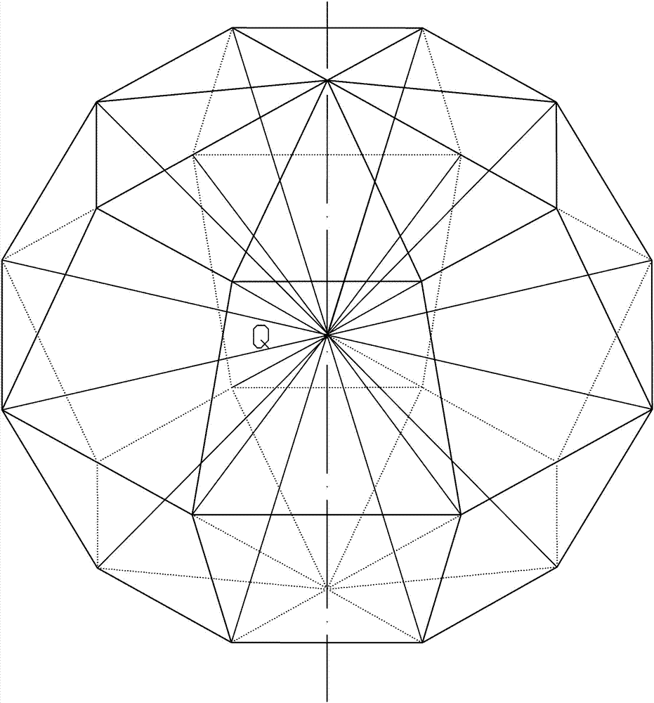
[0076] The theory is: assuming that object A and object B have the same volume (VA=VB), A is a perfect sphere, and B is an irregular shape, then the number of points of the smallest unit in object A and object B is the same, and these smallest units The straight-line distances formed by the points are equal. If any point in the cavity of object B is taken as the center point Q, and any point on the surface of B can be connected to the center point Q by a straight line, then the average value of the sum of the lengths of all radial lines passing through the center point in the irregular object must be equal to the diameter of A .

[0077] From this, it is deduced that for an irregular sh...

Embodiment 3

[0089] Such as Figure 8-9 , Figure 8 (a), (b), (c) and (d) are two-dimensional images of four sections of the bladder obtained by ultrasound during the specific implementation of the present invention.

[0090] The first step is to scan the bladder through the abdomen. The two-dimensional image of the bladder obtained by ultrasound, the display depth of the acoustic window is fixed, and the boundary of the bladder is clearly distinguished as much as possible. Try to show the mid-sagittal plane D1 of the bladder, that is, the long-axis section of the catheter or the long-axis section of the urethra, and the largest cross-section D2. The left oblique plane D3 and the right oblique plane D4 are obtained by rotating the probe clockwise and counterclockwise by about 45º respectively on the basis of the midsagittal plane. In principle, the above four cut planes are obtained by rotating the probe on the same axis.

[0091] The second step is the positioning method of Q point (th...


PUM


Abstract

The invention relates to a bladder volume measurement and calculation method under abdominal two-dimensional ultrasound. The method comprises the following steps that 1, the bladder is subjected to abdominal scanning, and a bladder two-dimensional image, namely, the median sagittal plane D1 is obtained through the ultrasound; 2, a probe is counterclockwise rotated by 90 degrees, and the maximum section, namely, the cross section D2 of the bladder median sagittal plane is obtained; 3, the probe is clockwise rotated by 45 degrees on the basis of the first median sagittal plane D1, and the maximum section, namely, the left slope D3 of the bladder median sagittal plane is obtained; 4, the probe is clockwise rotated by 45 degrees on the basis of the D2, and the maximum section, namely, the right slope D4 of the bladder median sagittal plane is obtained; 5, the center point Q of the median sagittal plane is positioned; 6, the median sagittal plane D1, the left slope D3 and the right slope D4 are measured; 7, the cross section D2 is measured; 8, the actual volume of the bladder is calculated according to a formula.

Description

technical field [0001] The invention relates to the field of bladder volume calculation methods, in particular to a bladder volume measurement and calculation method under transabdominal two-dimensional ultrasound. Background technique [0002] There are many methods for measuring and calculating bladder volume, such as CT, MRI, 131 I-OIH, ultrasound, catheterization, etc. Among them, catheterization is more accurate, and transabdominal ultrasound measurement is widely used in clinical practice due to its advantages of simple operation, low cost, non-invasiveness, and no damage. [0003] However, the shape of the bladder at different levels of filling is different. From very little residual volume to excessive filling volume, the shape of the sagittal plane changes greatly, and there is no obvious rule to follow. The shapes mainly include irregular narrow strips, irregular approximately small squares, irregular ovals, irregular trapezoids, approximately regular rounded squa...

Claims


Application Information

Patent Timeline
Patent Type & Authority Applications(China)
IPC IPC(8): A61B8/00
CPCA61B8/00A61B8/5223
Inventor 张茳全昆
Owner CHINA THREE GORGES UNIV
Why Eureka
Try Eureka