Medical image lesion area positioning and classification method

A technology of lesion area and medical image, applied in the field of image processing, can solve problems such as affecting the classification accuracy, and achieve the effect of improving the classification accuracy and improving the classification effect.

Inactive Publication Date: 2017-11-07
NANJING UNIV OF INFORMATION SCI & TECH
View PDF3 Cites 9 Cited by
  • Summary
  • Abstract
  • Description
  • Claims
  • Application Information

AI Technical Summary

Problems solved by technology

However, the existing intelligent analysis of medical images is mostly directly used to analyze the entire original image, and many of the extracted features have nothing to do with the lesion area, which seriously affects the classification accuracy.

Method used


Image

  • Medical image lesion area positioning and classification method
  • Medical image lesion area positioning and classification method
  • Medical image lesion area positioning and classification method

Examples


Embodiment Construction

[0023] The present invention will be described in detail below in conjunction with the accompanying drawings.

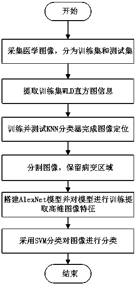
[0024] The method for locating and classifying the lesion area of ​​the medical image of the present invention automatically locates the lesion area first, then divides the image to retain the lesion area, and then trains the segmented image with a deep learning model, which can greatly improve the accuracy of classification. The flow chart of the invention is as follows figure 1 shown.

[0025] Specific steps are as follows:

[0026] 1) Extract WLD histogram features

[0027] For a given image, the WLD histogram features are extracted using differential excitation ξ and gradient direction θ.

[0028] A. Calculating differential incentives ξ

[0029] The sum of the grayscale difference between the center pixel and all its neighbor pixels is v 00 express:

[0030]

[0031] Among them, p represents the number of adjacent points, v i Represents the gray level o...


PUM


Abstract

The invention discloses a medical image lesion area positioning and classification method. The method comprises the following steps: the step 1, acquiring medical images, and dividing the images into a training set and a test set; the step 2, extracting WLD histogram information from the images of the training set, marking characteristic points of the training set according to the histogram information, placing the marked characteristic points into a KNN classifier, training the classifier, testing the trained KNN classifier using the images of the test set, and completing positioning of a medical image lesion area; the step 3, segmenting the images after the positioning through adoption of a histogram threshold method, and reserving the lesion area; and the step 4, putting the segmented images into a CNN depth model to extract characteristics, performing lesion area characteristic classification using an SVM classifier, and outputting a classification result. When the lesion area occupies a small part of the whole image, the accuracy rate is substantially improved.

Description

technical field [0001] The invention belongs to the field of image processing, and in particular relates to a method for locating and classifying lesion areas in medical images. Background technique [0002] In the medical field, most of the diseased areas on the patient's body are presented to the doctor through images, and the doctor judges the patient's condition and makes a diagnosis by analyzing the images. With the rapid development of computer technology, it is more and more closely integrated with medical imaging technology. Computers use intelligent algorithms to assist in the analysis of medical images, helping doctors to quickly analyze patients' conditions, reducing the workload of doctors and greatly improving diagnostic efficiency; With the development of medical care, computer-aided analysis of medical image technology is also integrated into it, helping patients and their family members to know the patient's condition at any time without leaving home. [000...

Claims


Application Information

Patent Timeline
Patent Type & Authority Applications(China)
IPC IPC(8): G06T7/00G06T7/11G06T7/73G06T7/44
CPCG06T7/0012G06T7/11G06T7/44G06T7/73G06T2207/20081G06T2207/20084G06T2207/30004
Inventor 张小瑞徐慧孙伟朱利丰宋爱国牛建伟
Owner NANJING UNIV OF INFORMATION SCI & TECH
Why Eureka
Try Eureka