A set of primers for detection of tussah microsporidia and their application

A technique for detecting microsporidia and primers, which is applied in the direction of microorganisms, microorganism-based methods, and microorganism measurement/inspection. Address technical limitations and reduce impact effects

Active Publication Date: 2021-02-12
辽宁省农业科学院大连生物技术研究所
View PDF5 Cites 0 Cited by
  • Summary
  • Abstract
  • Description
  • Claims
  • Application Information

AI Technical Summary

Benefits of technology

In summary, this new technique described in the previous patents allows researchers to quickly identify microscopes called Tsuumoviruses that cause diseases such as malaria. By combining these techniques together they have potential applications across various industries like food processing industry where there may occur large numbers of cases involving different types of bacteria circulating around them. These advances will make diagnosis faster and easier compared to traditional laboratory tests.

Problems solved by technology

This patented describes different methods for detecting tiny particles called Tsuwa Silwirl Microsporum (TSCM), also known as Microsphaera Colletotrichum L., commonly referred to as Cryptozoan Parasite, causing harmful effects upon livestock cropping systems due to nectroporation caused by parthenogeneans living inside them. Current techniques require expensive instrumentations and involve complex procedures like extractive culture followed by molecular diagnosis, making it challenging to apply these techniques effectively across various industries including agriculture, forestry, fishery, animal husbandry, and consumer products. Therefore, new solutions must address issues associated with current methods while maintaining productivity and quality standards.

Method used


Image

  • A set of primers for detection of tussah microsporidia and their application
  • A set of primers for detection of tussah microsporidia and their application
  • A set of primers for detection of tussah microsporidia and their application

Examples


Embodiment 1

[0066] Preparation of plasmid standards:

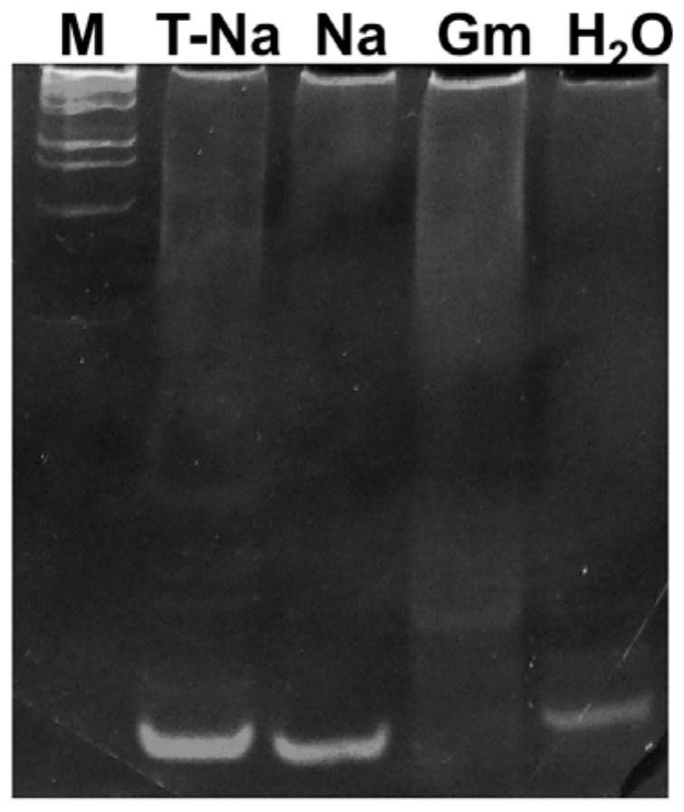
[0067] Take the tussah microsporidia suspension, centrifuge at 5000r / min for 5 minutes, remove the supernatant and retain the spore precipitate, add the extract (10mmol / L Tris-HCL pH 8.0; 10mmol / L EDTA pH 8.0; 100mmol / L NaCl; 2 %SDS; 0.039mol / L DTT), plus proteinase K (final concentration 100μg / mL), digested at 55°C for 4 hours, extracted with phenol / chloroform, washed twice with 70% ethanol, dried and eluted with TE to obtain Genomic DNA of Microsporidium tussah. The genomic DNA of the above-mentioned Microsporidia tussah was used for routine PCR. The PCR system was: 10×PCR buffer 2.5 μL, 10 mmol dNTPs 2 μL, MgCl2 2 μL, 10 μmol / L upstream and downstream primers 0.5 μL each, 1U Taq enzyme, template DNA 2 μL, add Sterile deionized water to 25 μL. Reaction conditions: pre-denaturation at 94°C for 5 minutes; 35 cycles at 94°C for 30s, 65°C for 30s, and 72°C for 60s; extension at 72°C for 10 minutes. After the reaction, take 10 μL of t...

Embodiment 2

[0079] Preparation of plasmid standards:

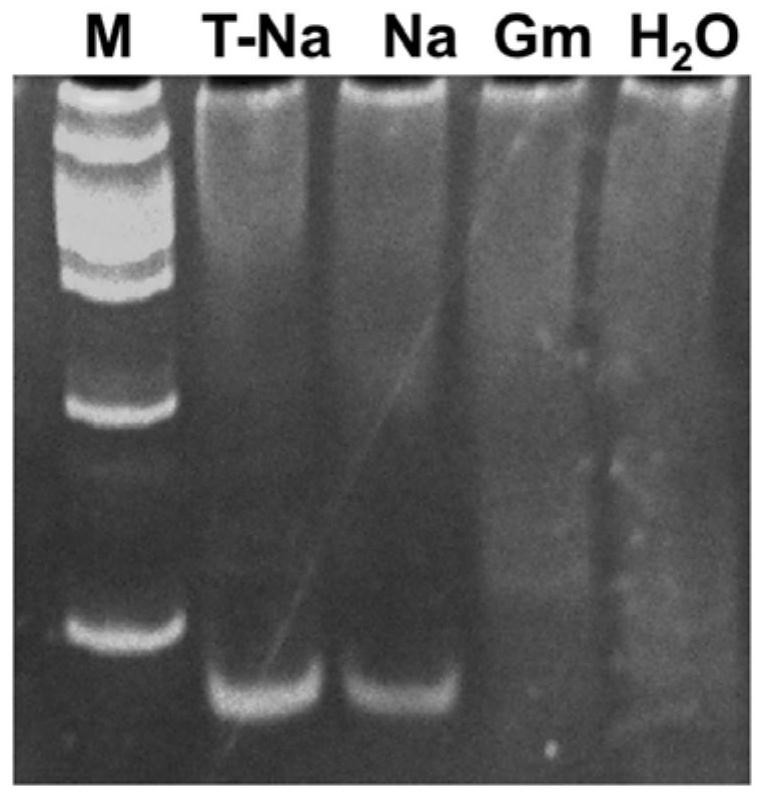
[0080]Take the tussah microsporidia suspension, centrifuge at 5000r / min for 5 minutes, remove the supernatant and retain the spore precipitate, add the extract (10mmol / L Tris-HCL pH 8.0; 10mmol / L EDTA pH 8.0; 100mmol / L NaCl; 2 %SDS; 0.039mol / L DTT), plus proteinase K (final concentration 100μg / mL), digested at 55°C for 4 hours, extracted with phenol / chloroform, washed twice with 70% ethanol, dried and eluted with TE to obtain Genomic DNA of Microsporidium tussah. The genomic DNA of the above-mentioned Microsporidia tussah was used for routine PCR. The PCR system was: 10×PCR buffer 2.5 μL, 10 mmol dNTPs 2 μL, MgCl2 2 μL, 10 μmol / L upstream and downstream primers 0.5 μL each, 1U Taq enzyme, template DNA 2 μL, add Sterile deionized water to 25 μL. Reaction conditions: pre-denaturation at 94°C for 5 minutes; 35 cycles at 94°C for 30s, 65°C for 30s, and 72°C for 60s; extension at 72°C for 10 minutes. After the reaction, take 10 μL of th...

Embodiment 3

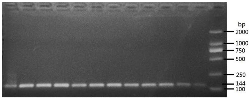
[0092] The DNA of Microsporidium tussah was automatically extracted by Thermo Nucleic Acid Automatic Extractor 706. A little tissue from 12 pupae with microparticle disease was taken, soaked in 210 μL of lysate (0.1M NaOH, 1% SDS) for 2 hours, and 200 μL of the supernatant was removed for use. Use the KF_TissueDNA_Duo program preset by the instrument to slightly adjust the amount of solvent in the DW wells of column A: take 200 μL of the lysate that has fully lysed the sample, 20 μL of the magnetic bead suspension (sigma), and 140 μL of absolute ethanol. Take 2 μL of the extracted DNA as a PCR template to configure a reaction system: each 25 μL system contains 0.5 μL of each primer (10 pM) shown in SEQ ID NO:4 and SEQ ID NO:5, 0.25 μL of rTaq, 2.5 μL of 10×Buffer, 2 μL of dNTPs, 17.25 μL of deionized water and 2 μL of DNA template. The PCR reaction conditions are 95°C for 4 minutes; 40 cycles of 95°C for 30 seconds, 65°C for 30 seconds, and 72°C for 1 minute, and then extende...


PUM


Abstract

The invention discloses a nosema pernyi detection primer and application thereof. Specifically, the base sequence of the primer is shown in SEQ ID NO. 1-3, and a template extraction method of the primer comprises the step of releasing, enrichment, separation and purification of DNA through alkaline lysis and magnetic beads. The primer is good in specificity and high in sensitivity; and according to a nosema pernyi detection method established through the primer, operation is simple and rapid, the cost is low, the flux is high, living pupa sampling can be achieved, the antijamming capability is high, the contamination risk is low, the quality of extracted DNA is high, and automatic extraction can be achieved. The method can be widely applied to molecular detection, the detection flux and the operation convenience can be remarkably improved, the false positive rate of detection can be obviously decreased, and the detection sensitivity and accuracy and the detection efficiency are improved. The nosema pernyi detection primer and the application thereof have important significance for centralized quarantine of antheraea pernyi granulosis and conservation of genetic resources in the antheraea pernyi industry.

Description


Claims


Application Information

Patent Timeline
Owner 辽宁省农业科学院大连生物技术研究所
Why Eureka
Try Eureka