Method and device for detecting and locating lesion in video-assisted thoracoscopy surgery

A positioning device and thoracoscopic technology, applied in the medical field, can solve problems such as inaccurate positioning and small lesions

Pending Publication Date: 2018-05-25
SUZHOU LANGKAI MEDICAL TECH
View PDF4 Cites 5 Cited by
  • Summary
  • Abstract
  • Description
  • Claims
  • Application Information

AI Technical Summary

Problems solved by technology

[0004] In order to solve the above technical problems, the embodiments of the present invention expect to provide a method and device for detecting and locating lesions in video-assisted thoracoscopic surgery, so as to solve the problem that the lesions cannot be accurately positioned due to their small size, softness, and depth.

Method used


Image

  • Method and device for detecting and locating lesion in video-assisted thoracoscopy surgery
  • Method and device for detecting and locating lesion in video-assisted thoracoscopy surgery
  • Method and device for detecting and locating lesion in video-assisted thoracoscopy surgery

Examples


Embodiment 10

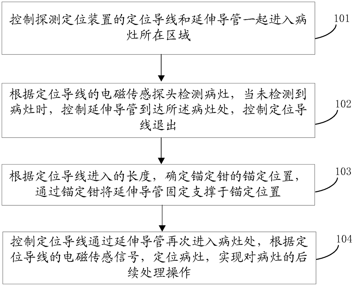
[0050] figure 1 A schematic flow chart of the lesion detection and positioning method in video-assisted thoracoscopic surgery provided by the embodiment of the present invention, as shown in figure 1 As shown, the executive body of this embodiment is the detection and positioning device, which includes: positioning wires, anchoring forceps, extension catheters and extracorporeal detectors. The detailed structure is described in the device part. In the embodiment of the present invention, the detection and positioning of lung cancer below 10 mm is taken as an example for illustration. The method includes:

[0051] Step 101: Control the positioning wire of the detection and positioning device and the extension catheter to enter the area where the lesion is located.

[0052] Step 102: Obtain lesion position information according to the positioning sensor of the positioning wire.

[0053] In steps 101 and 102, after the positioning wire of the control detection and positioning d...

Embodiment 2

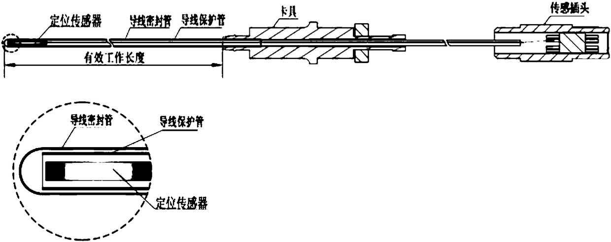
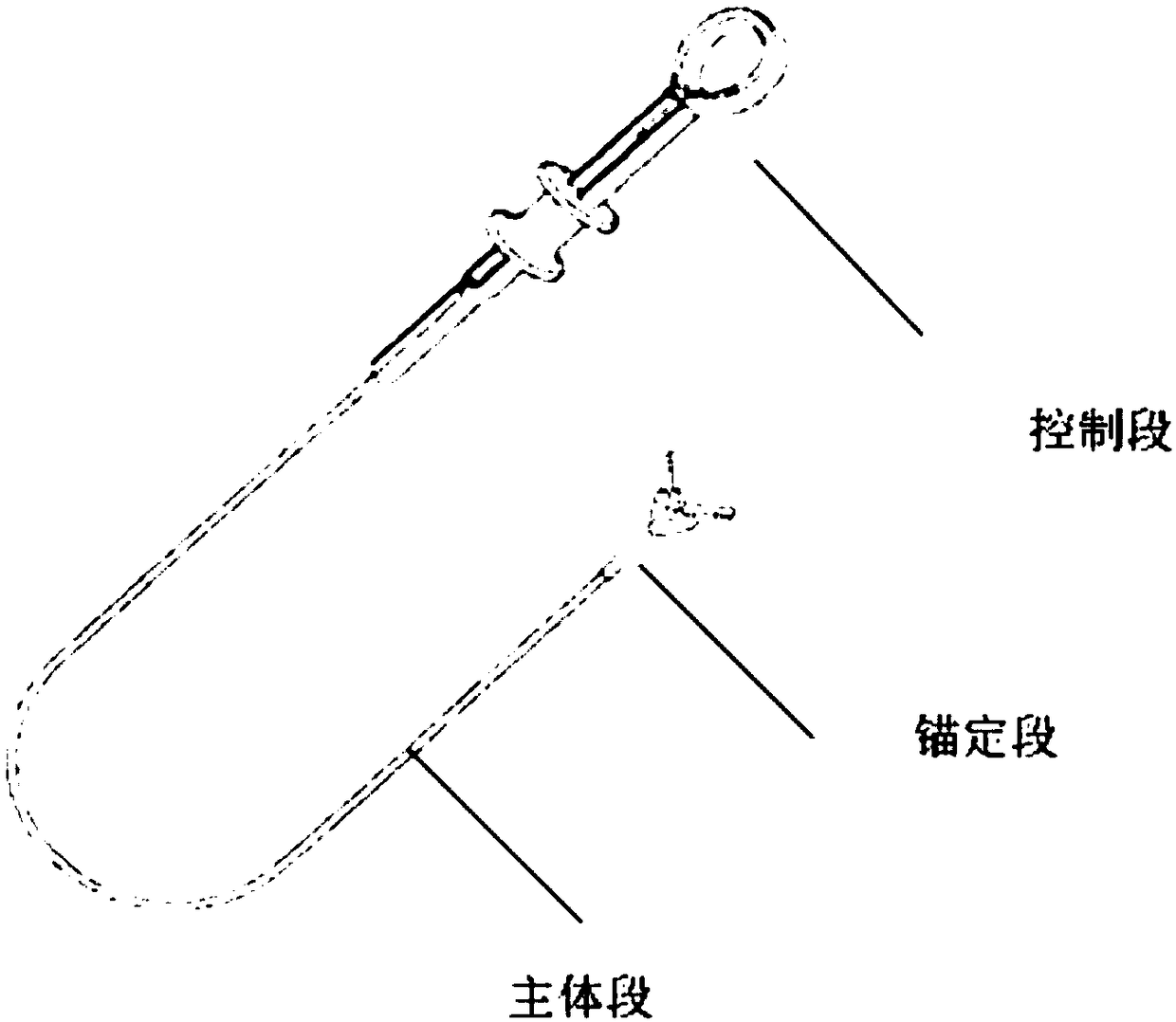
[0062] Figure 2A-2D Schematic diagram of the structure of the lesion detection and positioning device in video-assisted thoracoscopic surgery provided by the present invention, the device includes: positioning wires, anchoring forceps, extension catheters and external detectors; wherein, Figure 2A It is a schematic diagram of the structure of the positioning wire; Figure 2B is a schematic diagram of the structure of the anchor clamp; Figure 2C Schematic diagram of the structure of the extension catheter, Figure 2D Schematic diagram of the structure of the in vitro detector.

[0063] Wherein, the positioning wire is provided with a positioning sensor, and the positioning sensor is used to detect the current position coordinate information in the electromagnetic field; the positioning wire specifically includes:

[0064] Wire glands, wire protection tubes, positioning sensors, clamps and sensor plugs;

[0065] The wire sealing tube, the wire protection tube and the sensin...


PUM


Abstract

The embodiment of the invention discloses a method and device for detecting and locating a lesion in video-assisted thoracoscopy surgery. The method includes the steps that a locating wire with a locating sensor and an extension catheter together enter the area where the lesion is located; the location information of the lesion is detected through the locating sensor of the locating wire; the anchoring position of an anchoring tong is determined according to the length of the locating wire entering, and the extension catheter is fixed and supported at the anchoring position (the lesion position) through the anchoring tong; after a lung collapses in the process of surgery, the locating wire is controlled through the extension catheter to enter the lesion portion again, and the lesion is located through the locating sensor of the locating wire and an external detector used for detecting the position and the direction of the locating sensor. The method can locate the lesion location in real time, the location is more efficient and more accurate, and the problem of the difficulty in locating the lesion in VATS is solved.

Description

technical field [0001] The invention relates to the field of medical technology, in particular to a method and device for detecting and locating lesions in video-assisted thoracoscopic surgery. Background technique [0002] During video-assisted thoracic surgery (VATS) surgery, for currently common smaller, softer, deeper, intersegmental fissure, multiple, invisible, and intangible lesions, the lung collapses during the operation It is difficult to achieve intraoperative real-time localization of small lesions in the lung. [0003] The current existing solutions are not ideal, and it is difficult to achieve real-time intraoperative precise positioning. Preoperative positioning includes: transthoracic or transbronchopulmonary puncture methylene blue injection (diffusion), placement of microcoils (displacement), hook wire positioning (shedding), and radiotracer injection (radiation). Intraoperative positioning includes: intraoperative thoracoscopic B-ultrasound probe positio...

Claims


Application Information

Patent Timeline
Patent Type & Authority Applications(China)
IPC IPC(8): A61B90/11A61B34/20
CPCA61B34/20A61B90/11A61B2034/2051
Inventor 易新郑伟庄凌峰
Owner SUZHOU LANGKAI MEDICAL TECH
Why Eureka
Try Eureka