OCT fundus image semi-automatic segmentation method and device based on curve group matching

A fundus image, semi-automatic technology, applied in the field of medical image processing, can solve the problem of indistinct difference

Active Publication Date: 2020-05-05
SHANDONG NORMAL UNIV
View PDF0 Cites 0 Cited by
  • Summary
  • Abstract
  • Description
  • Claims
  • Application Information

AI Technical Summary

Problems solved by technology

[0005] In order to overcome the deficiencies of the above-mentioned prior art, the present invention combines dynamic time planning and multi-objective feature point matching technology, and proposes a new semi-automatic segmentation model based on curve group matching, which does not require any additional work. Under certain circumstances, any retinal layer in the fundus OCT image and the layered structure caused by some lesions can be accurately segmented, and it is not affected by the phenomenon that the difference between the layers of the central fovea in most images is not obvious

Method used


Image

  • OCT fundus image semi-automatic segmentation method and device based on curve group matching
  • OCT fundus image semi-automatic segmentation method and device based on curve group matching
  • OCT fundus image semi-automatic segmentation method and device based on curve group matching

Examples


Embodiment 1

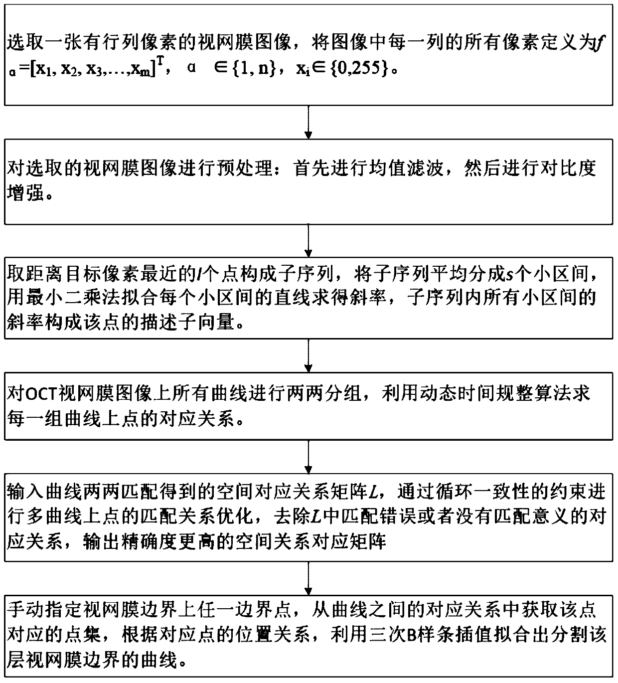
[0057] This embodiment discloses a semi-automatic retinal segmentation method, such as figure 1 shown, including the following steps:

[0058] Step 1: Select a retina image with m rows and n columns of pixels, and define all pixels in each column of the image as f α =[x 1 ,x 2 ,x 3 ..., x m ] T , α∈{1,n}, x i ∈{0,255}.

[0059] Step 2: Preprocessing the selected retinal image;

[0060] Step 2.1: Set a square sliding window with a size of 5×5 pixels, and perform mean value filtering on the image. The window starts to slide from the upper left corner of the image, and moves one pixel position each time, and calculates the mean value of 25 pixel values ​​in the window, and uses the mean value Replace the pixel value of the center point of the window, and this step is repeated until the window passes through all points on the image.

[0061] Step 2.2: Perform contrast enhancement on the filtered image, and calculate the median p of the gray value of each column of pixels ...

Embodiment 2

[0094] The purpose of this embodiment is to provide a computing device.

[0095] A semi-automatic segmentation device for OCT fundus images based on curve group matching, comprising a memory, a processor, and a computer program stored on the memory and operable on the processor, and the processor implements the following steps when executing the program, including:

[0096] Receive a fundus image with m rows and n columns and perform preprocessing;

[0097] Determine the gray value vector curve f of each column of the fundus image α =[x 1 ,x 2 ,x 3 ...,x m ] T , α∈{1,n}, x i ∈ {0,255}, calculate the gray value vector curve f α Descriptor of each pixel point in, obtain the descriptor matrix of the fundus image;

[0098] Based on the descriptor matrix, all gray value vector curves are pairwise matched through a dynamic time programming algorithm to obtain a spatial correspondence matrix between paired curve pixel points;

[0099] The pixel points manually specified by t...

Embodiment 3

[0101] The purpose of this embodiment is to provide a computer-readable storage medium.

[0102] A computer-readable storage medium, on which a computer program is stored, and when the program is executed by a processor, the following steps are performed:

[0103] Receive a fundus image with m rows and n columns and perform preprocessing;

[0104] Determine the gray value vector curve f of each column of the fundus image α =[x 1 ,x 2 ,x 3 ...,x m ] T , α∈{1,n}, x i ∈ {0,255}, calculate the gray value vector curve f α Descriptor of each pixel point in, obtain the descriptor matrix of the fundus image;

[0105] Based on the descriptor matrix, all gray value vector curves are pairwise matched through a dynamic time programming algorithm to obtain a spatial correspondence matrix between paired curve pixel points;

[0106] The pixel points manually specified by the user are received, and all pixel points corresponding to the points are found according to the coordinates of...


PUM


Abstract

The invention discloses an OCT eye fundus image semi-automatic segmentation method and device based on curve group matching. The method comprises: receiving and preprocessing an m-row n-column eye fundus image; determining the grayscale value vector curve of each column of the eye fundus image, calculating the descriptor of each pixel in the grayscale value vector curve, and obtaining a descriptorsub-matrix of the eye fundus image; and based on the description sub-matrix, matching every two grayscale value vector curves by using a dynamic time planning algorithm to obtain a spatial correspondence matrix between paired curved pixels; receiving a pixel manually designated by a user, searching out all pixels corresponding to the pixel according to the coordinate of the point and the spatialcorrespondence matrix, and fitting a smooth segmentation line. The technical solution of the invention ensures the segmentation accuracy of OCT eye fundus image, has strong adaptability, can segment the obvious layered structure on the retina, and can process the overlapping area at a central recess.

Description

technical field [0001] The invention relates to the field of medical image processing, in particular to a method and device for semi-automatic segmentation of OCT fundus images based on curve group matching. Background technique [0002] Optical Coherence Tomography (OCT) technology is the first microscopic medical imaging technology of biological tissue based on the principle of low-coherence light interference. Its theoretical basis is the early white light interferometry method. Because of its high resolution, non-invasive and non-invasive characteristics, it has great application value in the field of clinical medicine. Obtaining retinal images through OCT examination can clearly display the structure and function information of the fundus. A large number of studies have shown that changes in retinal nerve fiber layer (retinal never fiber layer, RNFL) thickness can be used as an important indicator of neurodegenerative diseases such as glaucoma and macular degeneration....

Claims


Application Information

Patent Timeline
Patent Type & Authority Patents(China)
IPC IPC(8): G06T7/10
CPCG06T7/10G06T2207/10101G06T2207/30041
Inventor 郑元杰段汶君连剑刘弘魏本征
Owner SHANDONG NORMAL UNIV
Why Eureka
Try Eureka