Method and device for processing eye fundus images

A fundus image and preprocessing technology, applied in the field of computer vision, can solve difficult diseases, difficult to apply massive fundus image processing, poor robustness and other problems

Active Publication Date: 2018-07-13
柯鑫
View PDF6 Cites 12 Cited by
  • Summary
  • Abstract
  • Description
  • Claims
  • Application Information

AI Technical Summary

Problems solved by technology

[0003] However, the differences in fundus images are very large due to different shooting equipment, technical level differences of shooting technicians, and differences in fundus of different patients. Applicable to various processing methods of different fundus images, that is to say, the robustness of processing and analyzing different types of fundus images in related technologies is poor, and it is difficult to apply to the processing of massive fundus images
At the same time, in related technologies, only general classification of fundus images can be made, and quantitative analysis of fundus images cannot be performed, making it difficult to accurately analyze related diseases of the fundus

Method used


Image

  • Method and device for processing eye fundus images
  • Method and device for processing eye fundus images
  • Method and device for processing eye fundus images

Examples


Embodiment Construction

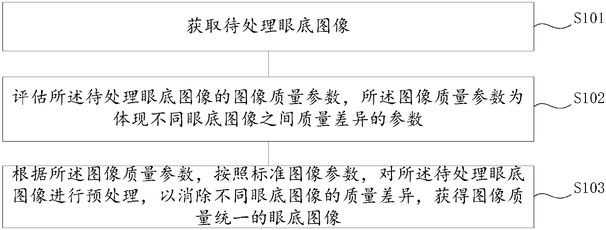
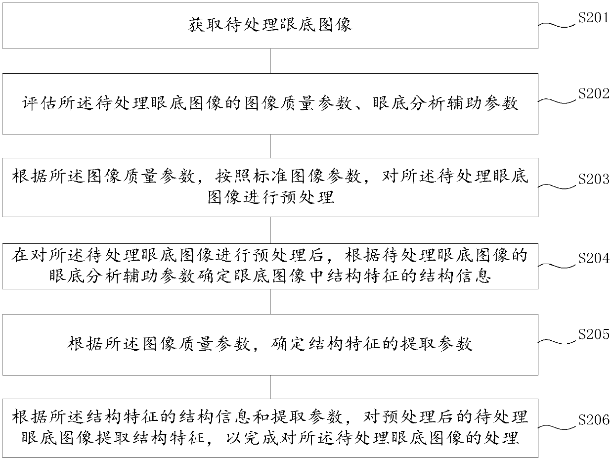
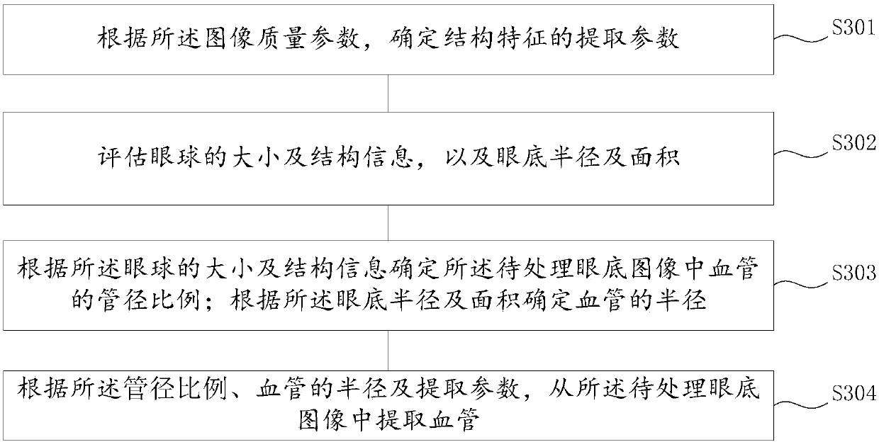
[0122] In order to make the purpose, technical solution and advantages of the present invention more clear, the embodiments of the present invention will be described in detail below in conjunction with the accompanying drawings. It should be noted that, in the case of no conflict, the embodiments in the present application and the features in the embodiments can be combined arbitrarily with each other.

[0123] The steps shown in the flowcharts of the figures may be performed in a computer system, such as a set of computer-executable instructions. Also, although a logical order is shown in the flowcharts, in some cases the steps shown or described may be performed in an order different from that shown or described herein.

[0124] The optic disc (English: optic disc), also known as the optic nerve head, is one of the important physiological and anatomical structures on the retina. It is a light red disc-shaped structure with clear boundaries, located about 3 mm from the nasal...


PUM


Abstract

The invention discloses a method and device for processing eye fundus images. The method includes the steps of obtaining to-be-processed eye fundus images; evaluating image quality parameters of the to-be-processed eye fundus images, wherein the image quality parameters are the parameters reflecting the quality differences among different eye fundus images; conducting preprocessing on the to-be-processed eye fundus images according to the image quality parameters and standard image parameters to remove the quality differences among different eye fundus images to obtain the eye fundus images with uniform image quality. By conducting preprocessing on the to-be-processed eye fundus images, not only are the quality differences among different eye fundus images removed, but also the calculationamount of structural feature extraction and lesion analysis in the later period is reduced, so that the running time for analyzing the to-be-processed eye fundus images is shortened, and the purposeof processing the great quantity of to-be-processed eye fundus images at the same time can be achieved. Finally, the accuracy of the structural feature extraction and lesion judgement is improved.

Description

technical field [0001] The invention relates to the technical field of computer vision, in particular to a method and device for processing fundus images. Background technique [0002] Studies have found that eye diseases and systemic diseases can be manifested in the retina to varying degrees. The systemic disease, such as stroke, hypertension, diabetes, cardiovascular disease (including coronary heart disease and cerebrovascular disease), the eye disease such as glaucoma, retinopathy caused by premature birth, papilledema, macular cleft and age-related macular degeneration. Therefore, through the processing of fundus images, the detection of eye diseases and systemic diseases has increasingly become the research focus in the field of computer vision. [0003] However, the differences in fundus images are very large due to different shooting equipment, technical level differences of shooting technicians, and fundus differences of different patients. It is applicable to v...

Claims


Application Information

Patent Timeline
IPC IPC(8): A61B3/12
CPCA61B3/12
Inventor 江威
Owner 柯鑫
Why Eureka
Try Eureka