Tissue excisor and tissue excising system
A resection and tissue technology, applied in sensors, parts of surgical instruments, medical science, etc., can solve problems such as uneconomical, patient danger, fatal injury, etc., and achieve the effect of increasing efficiency, reducing manufacturing costs, and being easy to replace.
- Summary
- Abstract
- Description
- Claims
- Application Information
AI Technical Summary
Problems solved by technology
Method used
Image
Examples
no. 1 approach
[0061] A tissue resection system equipped with a tissue resection device according to a first embodiment will be described below.
[0062] ●Tissue resection device
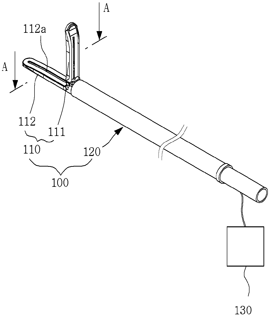
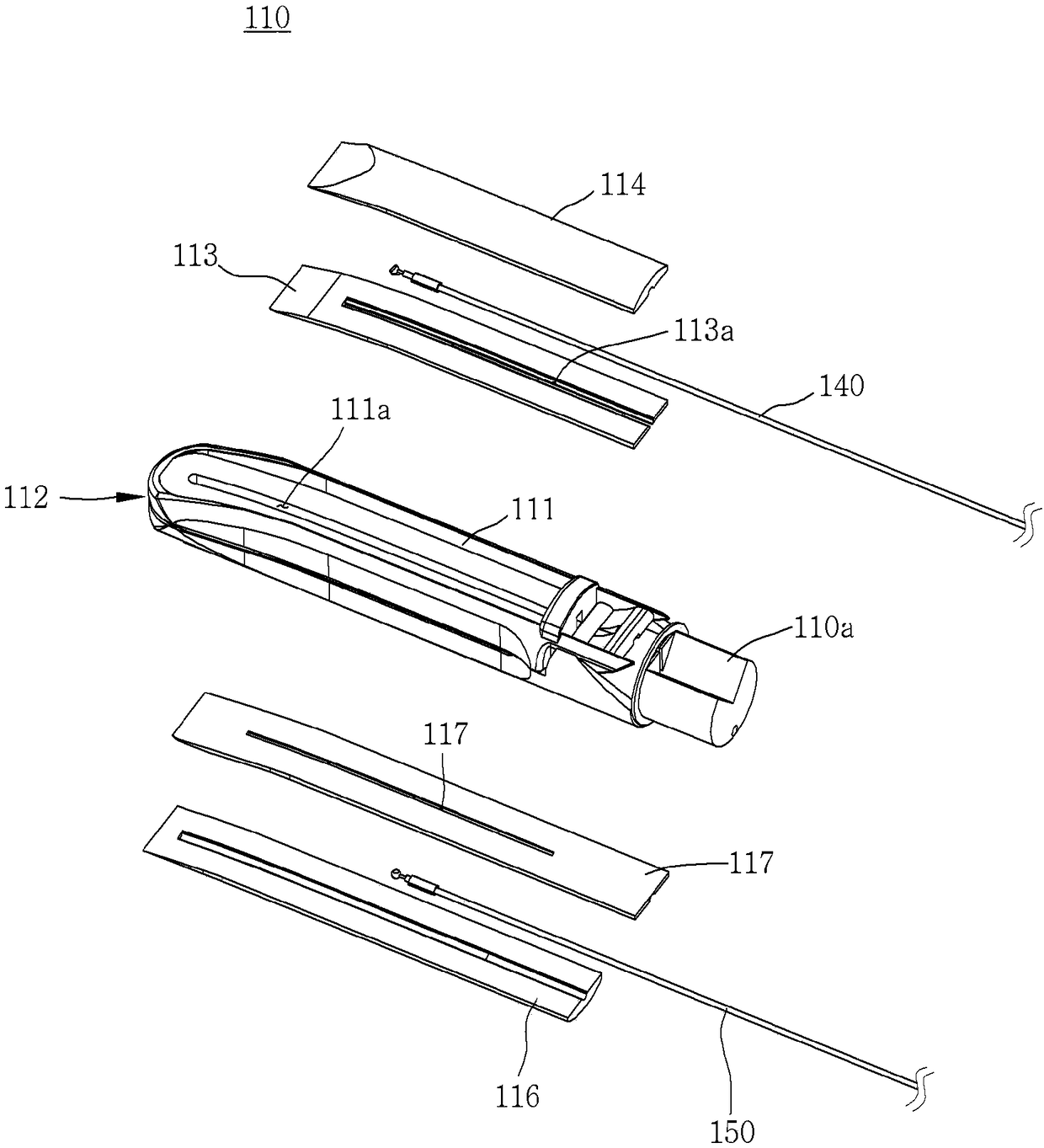
[0063] Such as figure 1 and figure 2 As shown, the tissue resection device 100 includes a resection unit 110 , an extension part 120 and an optical signal transmission module 140 .
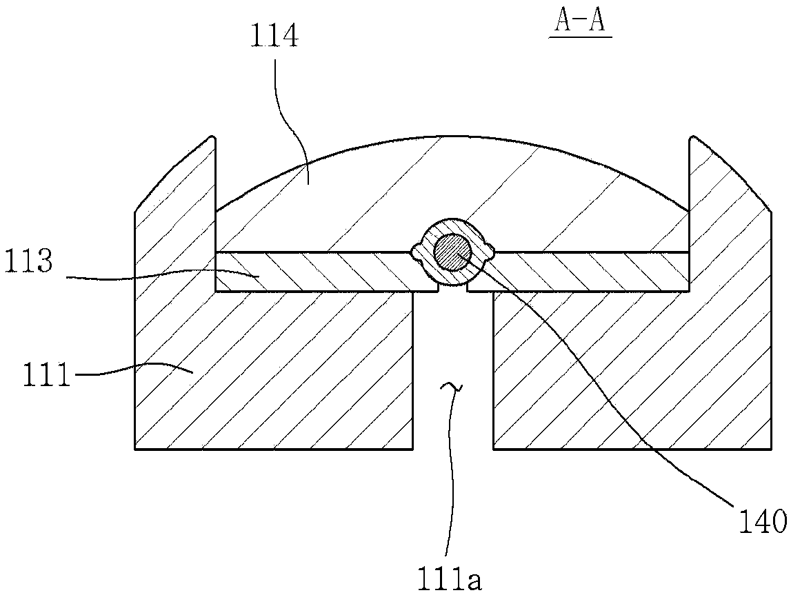
[0064] The cutting unit 110 is inserted into the human body to grasp or cut the human tissue 10 . An optical signal transmission module 140 for line-scanning a portion to be resected from the human tissue 10 is provided in the resection unit 110 . A second optical signal transmission module 150 operating independently of the optical signal transmission module 140 may also be provided in the cutting unit 110 .
[0065] The optical signal transmission module 140 and the second optical signal transmission module 150 may be configured to provide a reflected optical signal or a reflected second optical signal to the image generation u...
no. 2 approach
[0156] A tissue resection device 100a and a tissue resection system 200a using the tissue resection device 100a according to a second embodiment of the present invention are described below.
[0157] The tissue resection device 100a according to the present embodiment is the same as the first embodiment described above in terms of structures and functions of the resection unit 110 , the extension 120 , and the actuator 130 . In addition, the installation positions and structures of the optical signal transmission module 140a and the second optical signal transmission module 150a are the same as those of the optical signal transmission module 140 and the second optical signal transmission module 150 of the above-mentioned first embodiment, therefore, the following description of the optical signal transmission module 140a and the second optical signal transmission module 150a are different from the first embodiment.
[0158] In this embodiment, the optical signal transmission m...
no. 3 approach
[0166] Refer below Figure 15 to Figure 19 A tissue resection device 300 according to a third embodiment of the present invention and a tissue resection system 200b using the same will be described.
[0167] Such as Figure 15 to Figure 18 As shown, the tissue resection device 300 according to this embodiment includes a resection unit 310, an extension part 320, an actuator 330, a pair of optical signal transmission modules 340a and 340b, and a pair of second optical signal transmission modules.
[0168] The cutting unit 310 , the extension part 320 and the actuator 330 of the present embodiment basically have the same functions as the cutting unit 110 , the extension part 120 and the actuator 120 of the first embodiment described above. In addition, the pair of optical signal transmission modules 340a and 340b and the pair of second optical signal transmission modules are functionally and structurally the same as the optical signal transmission module 140 and the second opti...
PUM
 Abstract
Description
Claims
Application Information
 - R&D
- Intellectual Property
- Life Sciences
- Materials
- Tech Scout
- Unparalleled Data Quality
- Higher Quality Content
- 60% Fewer Hallucinations
Browse by: Latest US Patents, China's latest patents, Technical Efficacy Thesaurus, Application Domain, Technology Topic, Popular Technical Reports.
© 2025 PatSnap. All rights reserved.Legal|Privacy policy|Modern Slavery Act Transparency Statement|Sitemap|About US| Contact US: help@patsnap.com


