
Biosensor for detecting transport, and application of biosensor

A technology for biosensors and object measurement, applied in the field of biosensors for detecting translocation

Active Publication Date: 2018-08-10
EAST CHINA NORMAL UNIV +1
View PDF2 Cites 0 Cited by
  • Summary
  • Abstract
  • Description
  • Claims
  • Application Information

AI Technical Summary

Problems solved by technology

[0005] However, there is currently a lack of satisfactory methods for the efficient and accurate detection of biotransport functions in vitro

Method used


Image

  • Biosensor for detecting transport, and application of biosensor
  • Biosensor for detecting transport, and application of biosensor
  • Biosensor for detecting transport, and application of biosensor

Examples


Embodiment 1

[0107] Preparation of biosensors

[0108] In this example, a biosensor was prepared based on mouse crypts, the method is as follows:

[0109] (1) 8-10 weeks old C57BL / 6 mouse CO 2 After euthanasia, the small intestines were dissected out and placed in pre-cooled PBS.

[0110] (2) Use dissecting forceps to carefully remove the fat tissue on the outer wall of the small intestine, then use ophthalmic scissors to open the small intestine cavity longitudinally, wash it with pre-cooled PBS for 5 times, transfer it to a sterile 50mL centrifuge tube, and place it on ice.

[0111] (3) In a sterile ultra-clean bench, wash the small intestine again 5 times with pre-cooled PBS containing penicillin / streptomycin (P / S).

[0112] (4) Transfer it to 50 mL of PBS containing 2 mM EDTA, and place it in a refrigerator at 4°C for 25 minutes for digestion.

[0113] (5) Transfer the digested small intestine to a 50mL centrifuge tube containing 25mL pre-cooled PBS, shake it back and forth about 50...

Embodiment 2

[0122] Observation of Biosensor Morphology

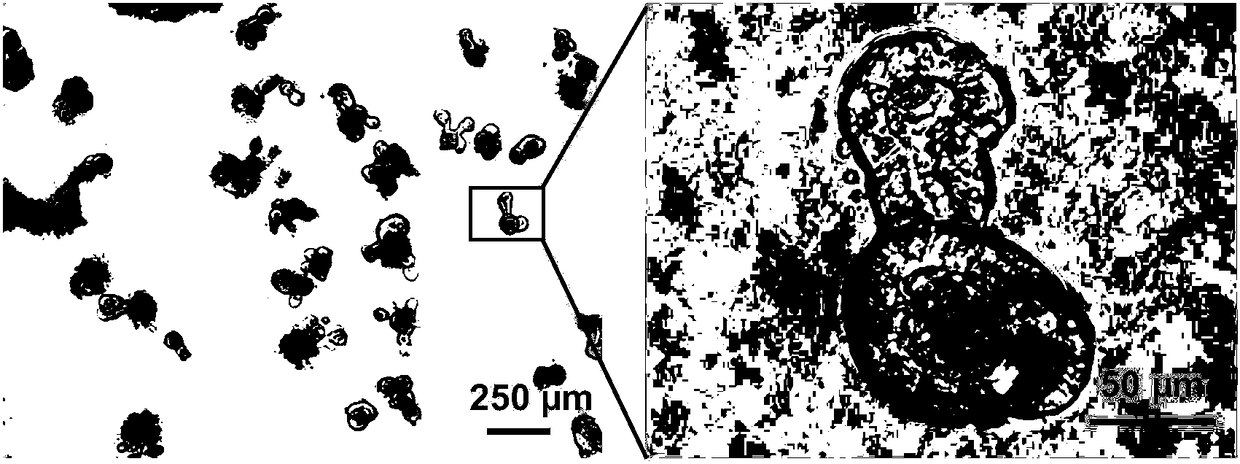
[0123] In this example, the morphology of the biosensor prepared in Example 1 was observed, wherein an Olympus DP 71 inverted microscope was used to observe and photograph the morphology of the biosensor cultured for 3 days.

[0124] The result is as figure 1 As shown, isolated crypts differentiated to form biosensors after 3 days in culture, and stem cells in the crypts began to differentiate to "bud out" to form new crypts.

Embodiment 3

[0126] Biosensor with MRP2 transport function

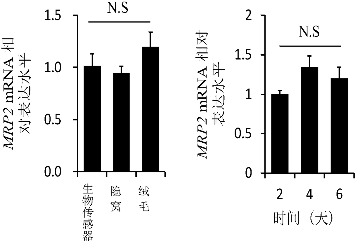
[0127] In this example, it is indirectly proved that the biosensor prepared in Example 1 has MRP2 transport function through the detection of mRNA expression level. Methods as below:

[0128] For the biosensor prepared in Example 1, the differentiation was induced in vitro for different days, the culture medium was discarded, washed once with pre-cooled PBS, and the matrigel was broken with a pipette tip, and transferred to a 1.5 mL container with pre-cooled PBS. In a centrifuge tube, centrifuge at 200g for 5 minutes at 4°C, discard the supernatant, and collect the biosensor. For the collected biosensors, total RNA was extracted by Trizol, and cDNA was synthesized by reverse transcription. Using the synthesized cDNA as a template, real-time quantitative PCP was used to analyze the expression level of MRP2 in the biosensor differentiated in vitro.

[0129] Result: if figure 2 As shown, the MRP2 mRNA expression levels of the i...


PUM


Abstract

The invention provides a biosensor for detecting transport, and application of the biosensor. Specifically, a method provided by the invention comprises the following steps: (a) providing a detectionsystem, wherein the detection system comprises (a1) a biosensor; (a2) a detection solution; (b) mixing the detection system with a substance to be detected and a transport inhibitor, or mixing the detection system with the substance to be detected and a transport substrate so as to form a first mixed solution; (c) optionally placing the first mixed solution for a period of time t; (d) separating the biosensor from the first mixed solution to obtain a separated biosensor; (e) detecting to obtain a transport detection result of the substance to be detected, or obtain a result of influence of thesubstance to be detected on a transporter. The biosensor is simple and convenient to operate as well as efficient and rapid in detection.

Description

technical field [0001] The invention belongs to the field of biotechnology, in particular to a biosensor for detecting translocation and its application. Background technique [0002] The transport of substances is related to many physiological activities. The ATP-binding cassette transporter is a type of superfamily transporter that has been studied more at present. It consists of two transmembrane domains and two cytoplasmic ATP-binding domains. It has a wide range of substrates and participates in bile salts, nucleosides, cholesterol, peptides , the transport of various endogenous and exogenous substrates such as various drugs, chloride ions, toxins, organic anions, iron, and sterols. [0003] At present, 49 human ABC transporter members have been found, which are divided into seven subfamilies (ABC-A to ABC-G). Some efflux transporters are often closely related to the drug resistance of many drugs. Therefore, ABC transporters are considered to be important targets for ...

Claims


Application Information

Patent Timeline
Patent Type & Authority Applications(China)
IPC IPC(8): G01N33/68G01N33/15
CPCG01N33/15G01N33/68
Inventor 王昕张磊张远金刘明耀席在喜
Owner EAST CHINA NORMAL UNIV
Learn More