
Blood vessel endoscopic imaging probe and imaging method

A technology for imaging probes and blood vessels, applied in endoscopes, catheters, ultrasonic/acoustic/infrasonic image/data processing, etc., can solve the problems of insufficient detection depth, low resolution, poor imaging quality, etc. Effect of Large Detection Depth

Pending Publication Date: 2018-08-21
武汉艾欧医疗科技有限公司
View PDF10 Cites 0 Cited by
  • Summary
  • Abstract
  • Description
  • Claims
  • Application Information

AI Technical Summary

Problems solved by technology

OCT has high imaging resolution and good imaging quality, but the detection depth is not enough, it can only detect the surface of the imaging part, and cannot detect the deep part of the imaging part
On the contrary, IVUS can be used for deep detection of imaging parts, but its resolution is low and the imaging quality is poor

Method used


Image

  • Blood vessel endoscopic imaging probe and imaging method
  • Blood vessel endoscopic imaging probe and imaging method
  • Blood vessel endoscopic imaging probe and imaging method

Examples


Embodiment Construction

[0030] The principles and features of the present invention are described below in conjunction with the accompanying drawings, and the examples given are only used to explain the present invention, and are not intended to limit the scope of the present invention.

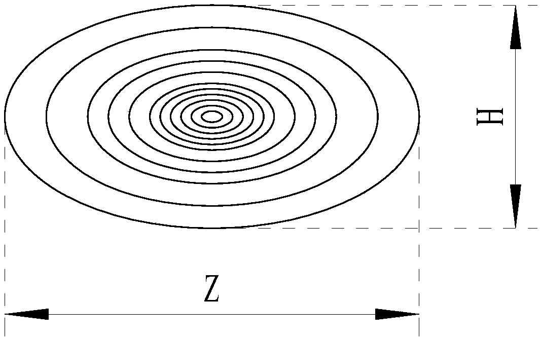
[0031] Because part of the optical path of the optical probe of the traditional photoacoustic endoscopy system is not focused, the projected spot of the laser emitted from the optical probe on the vessel wall is elliptical, with the strongest light intensity in the center and gradually weakens to the surrounding. Such as figure 1 As shown, the length of the light spot distribution along the axial direction is Z, the length of the light spot distribution along the radial direction is H, and Z is always greater than H, that is, the optical path distribution range in the axial direction is larger than that in the radial direction.

[0032] Since the sound wave range of the ultrasonic transducer is fixed, the greater th...


PUM


Abstract

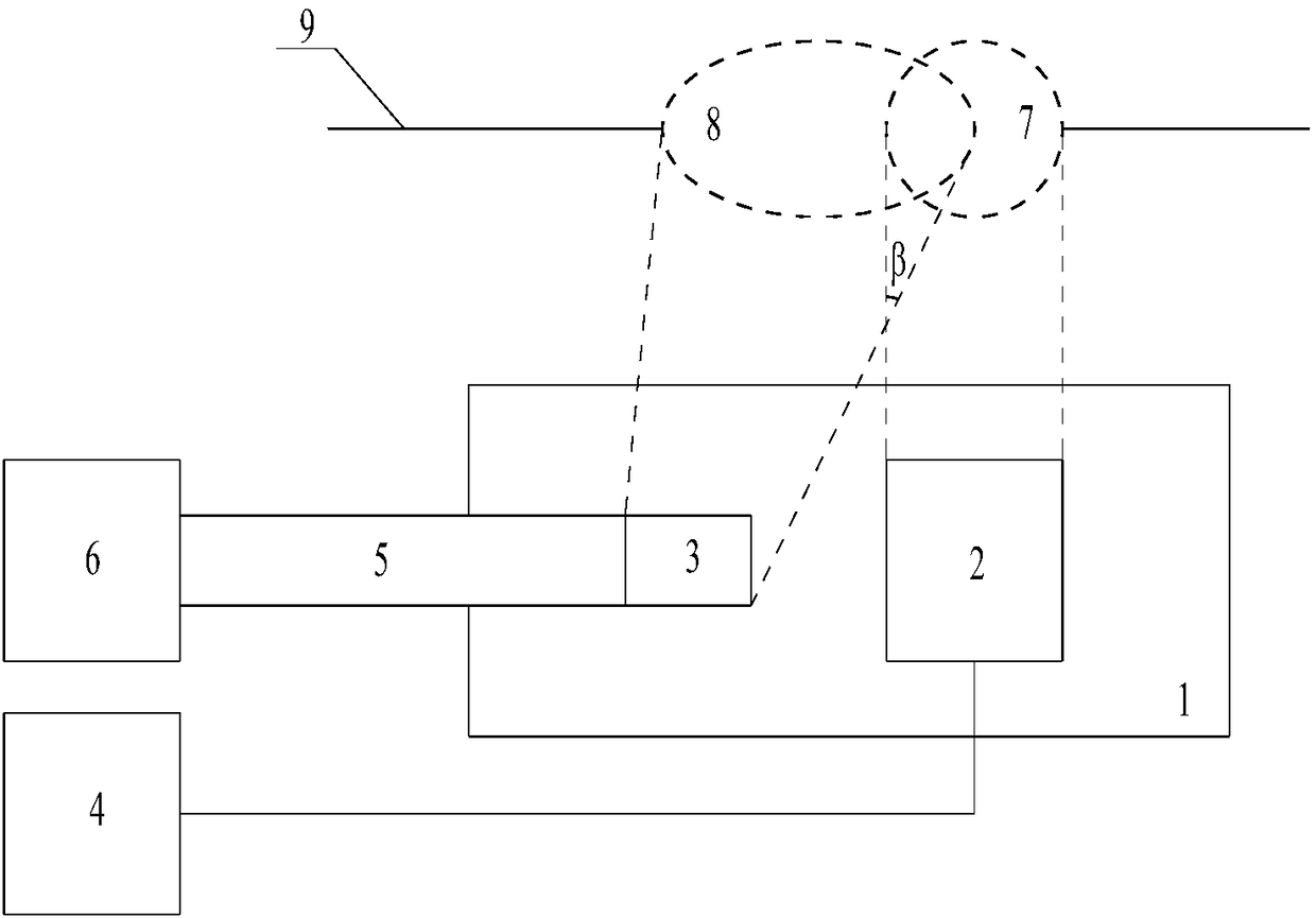
The invention relates to a blood vessel endoscopic imaging probe and imaging method. The probe includes a cylindrical casing. An ultrasonic transducer and a prism are sequentially arranged in the axial direction in the cylindrical casing, the ultrasonic transducer is electrically connected with a signal processor, the prism is connected to a laser through a multi-mode fiber matched with the prism,an ultrasonic projection area of the ultrasonic transducer on the blood vessel wall and a laser projection area of the prism on the blood vessel wall coincide, and the projecting line direction fromthe ultrasonic transducer to the ultrasonic projection area and the projecting line direction from the prism to the laser projection area form an included angle. According to the technical scheme, theprobing depth and imaging quality of the blood vessel endoscopic imaging probe can be both considered.

Description

technical field [0001] The invention relates to the technical field of medical devices, in particular to a blood vessel endoscopic imaging probe and an imaging method. Background technique [0002] Cardiovascular and cerebrovascular diseases have increasingly become one of the main diseases that seriously affect people's healthy life. According to statistics, tens of millions of people die of cardiovascular and cerebrovascular diseases in the world every year. The main cause of cardiovascular and cerebrovascular diseases is the sudden rupture of vulnerable plaques growing in atherosclerotic blood vessels, causing blockage of arteries, which interrupts blood supply to blood vessels and ultimately endangers life. [0003] At present, before the diagnosis of cardiovascular and cerebrovascular diseases in medicine, it is usually necessary to observe the vulnerable plaques in the cardiovascular and cerebrovascular through probes and imaging methods, and use the observed images as...

Claims


Application Information

Patent Timeline
Patent Type & Authority Applications(China)
IPC IPC(8): A61B8/12A61B8/08A61B5/00A61B1/00A61B1/06
CPCA61B1/00165A61B1/0661A61B5/0033A61B5/0084A61B5/0095A61B8/0891A61B8/12A61B8/5261
Inventor 魏伟王自昱
Owner 武汉艾欧医疗科技有限公司
Learn More