Placement device for visual ventricle and abdominal cavity shunt tube and application method thereof

A technology of shunt tube and abdominal cavity, which is applied in the medical field of implantation, which can solve the problems of abdominal shunt cut, insertion into the intestine, and danger to patients, and achieve high accuracy, high success rate, and avoid insertion errors.

Inactive Publication Date: 2018-09-11
LISHUI PEOPLES HOSPITAL
View PDF9 Cites 0 Cited by
  • Summary
  • Abstract
  • Description
  • Claims
  • Application Information

AI Technical Summary

Problems solved by technology

In recent years, ventriculoperitoneal shunts have been placed laparoscopically, avoiding the above-mentioned disadvantages such as large trauma, but the ventriculoperitoneal shunts must be inserted into the abdominal cavity with a trocar with a blunt head, and then the proximal end of the ventriculoperitoneal shunt must be inserted subcutaneously through the abdomen to the head and neck. It is connected to the ventricular end of the ventriculoperitoneal shunt tube. During this process, the peritoneal shunt tube is likely to be cut by a blunt instrument, causing danger to the patient, and the insertion is to enter the gap between the intestine and the intestinal wall. The existing insertion method is Insert in a straight line, if the operation is not good, the tube will be inserted directly into the intestine, causing damage

Method used


Image

  • Placement device for visual ventricle and abdominal cavity shunt tube and application method thereof
  • Placement device for visual ventricle and abdominal cavity shunt tube and application method thereof
  • Placement device for visual ventricle and abdominal cavity shunt tube and application method thereof

Examples


Embodiment 1

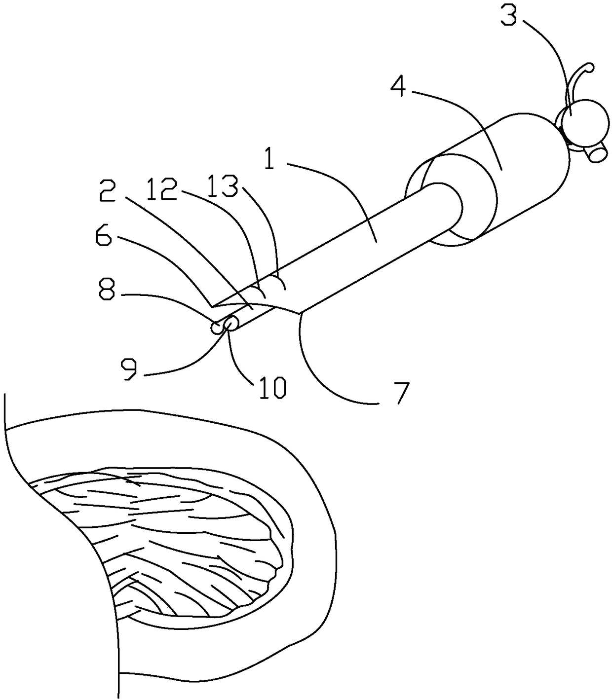
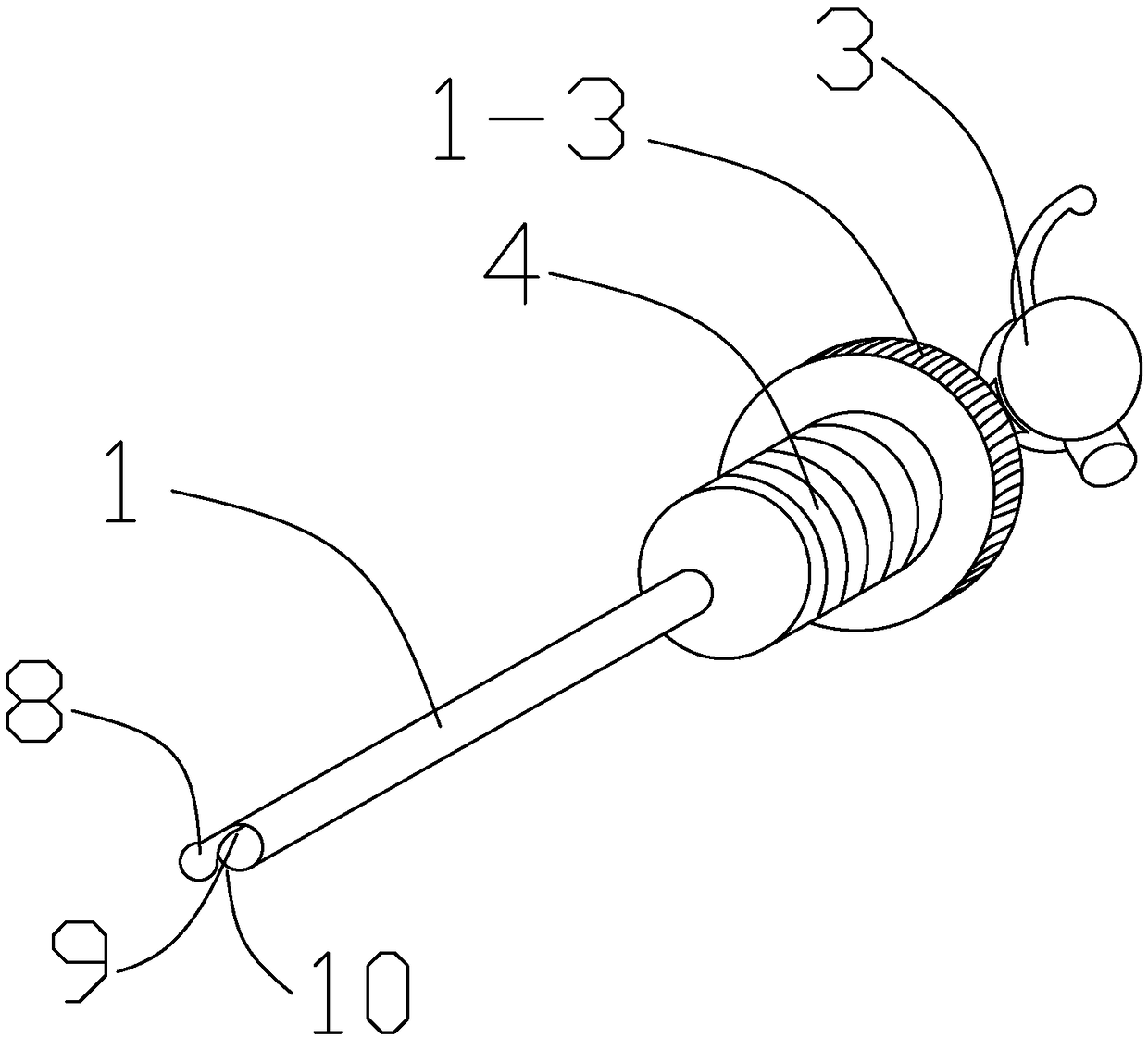
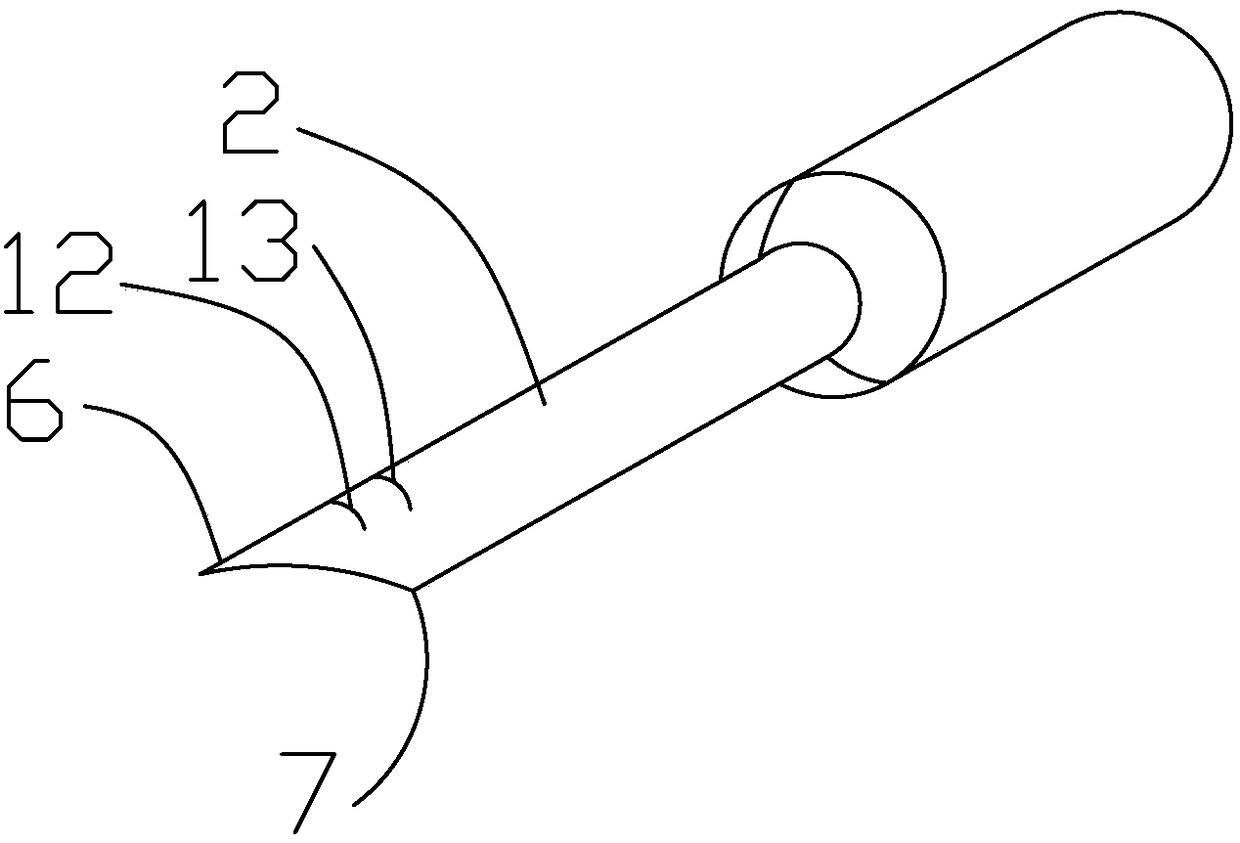
[0024] A device for placing a visualized ventriculoperitoneal shunt tube, comprising an inner cannula 1 and a sheath 2, the rear end of the inner cannula 1 is connected with a handle 3, the inner cannula 1 is provided with a spring 4, and the interior of the inner cannula 1 is A cavity, the inner sleeve 1 between the spring 4 and the handle 3 is also equipped with a first screw 1-2, the sheath 2 is installed on the outside of the inner sleeve 1, and the front end of the inner sleeve 1 protrudes from the sheath 2, the sheath 2 is also provided with a second threaded part 1-3 that matches the first threaded part 1-2, and is fixed to each other by the first threaded part 1-2 and the second threaded part 1-3. The front end of the sheath 2 is an inclined surface, the protruding part of the inclined surface is an outward sharp point 6, the other part of the inclined surface is an inner blunt point 7, the front end of the inner sleeve 1 It is a blunt head 8, the rear end surface of t...

Embodiment 2

[0027] A device for placing a visualized ventriculoperitoneal shunt tube, comprising an inner cannula 1 and a sheath 2, the rear end of the inner cannula 1 is connected with a handle 3, the inner cannula 1 is provided with a spring 4, and the interior of the inner cannula 1 is A cavity, the inner sleeve 1 between the spring 4 and the handle 3 is also equipped with a first screw 1-2, the sheath 2 is installed on the outside of the inner sleeve 1, and the front end of the inner sleeve 1 protrudes from the sheath 2, the sheath 2 is also provided with a second threaded part 1-3 that matches the first threaded part 1-2, and is fixed to each other by the first threaded part 1-2 and the second threaded part 1-3. The front end of the sheath 2 is an inclined surface, the protruding part of the inclined surface is an outward sharp point 6, the other part of the inclined surface is an inner blunt point 7, the front end of the inner sleeve 1 It is a blunt head 8, the rear end surface of t...

Embodiment 3

[0030] A device for placing a visualized ventriculoperitoneal shunt tube, comprising an inner cannula 1 and a sheath 2, the rear end of the inner cannula 1 is connected with a handle 3, the inner cannula 1 is provided with a spring 4, and the interior of the inner cannula 1 is A cavity, the inner sleeve 1 between the spring 4 and the handle 3 is also equipped with a first screw 1-2, the sheath 2 is installed on the outside of the inner sleeve 1, and the front end of the inner sleeve 1 protrudes from the sheath 2, the sheath 2 is also provided with a second threaded part 1-3 that matches the first threaded part 1-2, and is fixed to each other by the first threaded part 1-2 and the second threaded part 1-3. The front end of the sheath 2 is an inclined surface, the protruding part of the inclined surface is an outward sharp point 6, the other part of the inclined surface is an inner blunt point 7, the front end of the inner sleeve 1 It is a blunt head 8, the rear end surface of t...


PUM


Abstract

The invention belongs to the field of implantable medical treatment and particularly relates to an implantable auxiliary instrument for a visual ventricle and abdominal cavity shunt tube. A placementdevice for the visual ventricle and abdominal cavity shunt tube comprises an inner cannula and a sheath, a handle is connected to the rear end of the inner cannula, and a spring is arranged on the inner cannula; a cavity is formed in the inner cannula, a first threaded part is also installed on the inner cannula between the spring and the handle, a blunt end for integrated protection is added, a blunt instrument point can be prevented from scratching the abdominal cavity shunt tube, and unnecessary emergency conditions in the surgery are caused; through arrangement of a micro camera, minimallyinvasive surgery in ventriculo-peritoneal shunt can be observed, the blunt instrument point can also be prevented from scratching the abdominal cavity shunt tube, the unnecessary emergency conditionsin the surgery are caused, the insertion depth can also be observed, and the device helps a user to more accurately and conveniently implement the operation.

Description

technical field [0001] The invention belongs to the field of medical implantation, in particular to the implantation assistance of a ventriculoperitoneal shunt tube. Background technique [0002] For ventriculoperitoneal shunt surgery, when the abdominal end of the ventriculoperitoneal shunt tube is placed in the abdominal cavity, it must be placed through a laparotomy, which is traumatic and can easily lead to complications of laparotomy. In recent years, ventriculoperitoneal shunts have been placed laparoscopically, avoiding the above-mentioned disadvantages such as large trauma, but the ventriculoperitoneal shunts must be inserted into the abdominal cavity with a trocar with a blunt head, and then the proximal end of the ventriculoperitoneal shunt must be inserted subcutaneously through the abdomen to the head and neck. It is connected to the ventricular end of the ventriculoperitoneal shunt tube. During this process, the peritoneal shunt tube is likely to be cut by a blu...

Claims


Application Information

Patent Timeline
Patent Type & Authority Applications(China)
IPC IPC(8): A61B17/34
CPCA61B17/3415A61B17/3421A61B17/3494A61B2017/3425A61B2017/3456
Inventor 刘进黄刚江银华吴凌慧叶碎林程林贵梅明荣蓝威徐宇
Owner LISHUI PEOPLES HOSPITAL
Why Eureka
Try Eureka