Endoscopic surgery glasses-free 3D image display system and method

An image display and endoscope technology, applied in the field of naked-eye 3D image display system for endoscopic surgery, can solve problems such as inconvenient switching and affecting surgical safety, and achieve the effect of reducing burden and improving surgical safety

Pending Publication Date: 2018-09-21
ZHANGJIAGANG KANGDE XIN OPTRONICS MATERIAL
View PDF7 Cites 9 Cited by
  • Summary
  • Abstract
  • Description
  • Claims
  • Application Information

AI Technical Summary

Problems solved by technology

However, when using the traditional polarized 3D system, users need to wear glasses. Due to the long operation time, it will bring additional burden to doctors, especially those who wear myopia glasses.
At the same time, during the operation, when multiple doctors operate at the same time, it is inconvenient to switch between the endoscopic screen and the external scene, and even affect the safety of the operation.

Method used


Image

  • Endoscopic surgery glasses-free 3D image display system and method
  • Endoscopic surgery glasses-free 3D image display system and method
  • Endoscopic surgery glasses-free 3D image display system and method

Examples


Embodiment 1

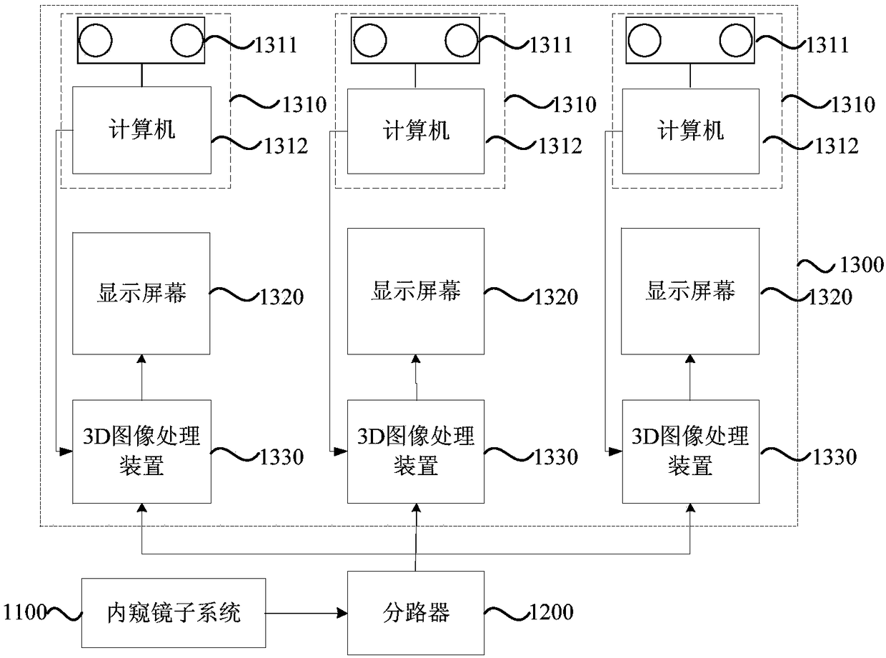
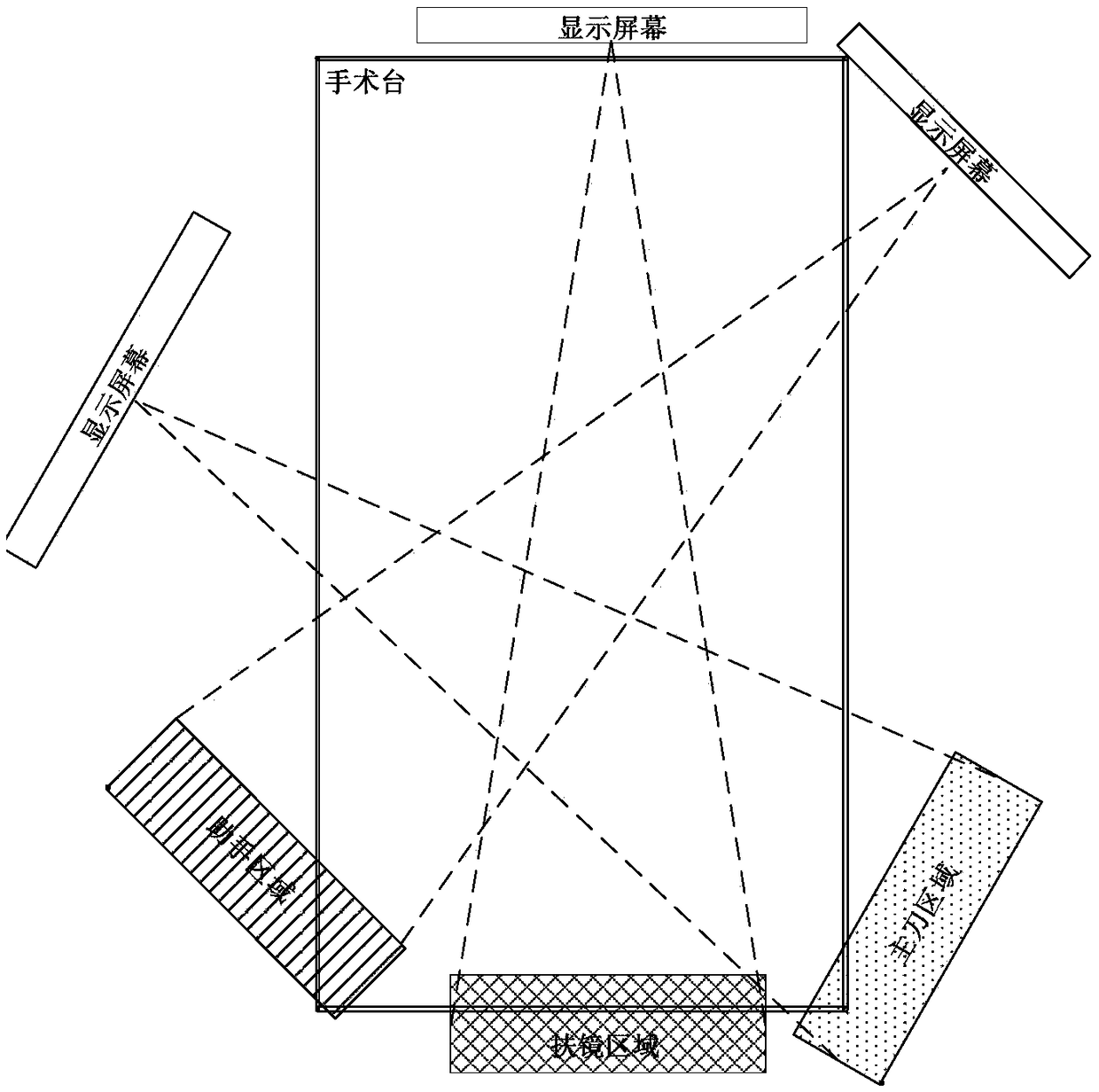
[0027] figure 1 It is a schematic structural diagram of a naked-eye 3D image display system for endoscopic surgery provided by Embodiment 1 of the present invention. This embodiment is applicable to viewing 3D surgical object images during endoscopic surgery. In this embodiment, according to the conventional configuration of surgical personnel, taking the chief surgeon, assistant doctor and mirror doctor participating in endoscopic surgery as an example, three naked-eye 3D image display subsystems are set in the system, such as figure 1 As shown, the system specifically includes:

[0028] An endoscope system 1100, a splitter 1200, and at least one naked-eye 3D image display subsystem 1300;

[0029] Wherein, the endoscope system 1100 is respectively connected with each naked-eye 3D image display subsystem 1300 through the splitter 1200, and is used to capture the image of the surgical object, and send the surgical object image to each naked-eye 3D image through the splitter 12...

Embodiment 2

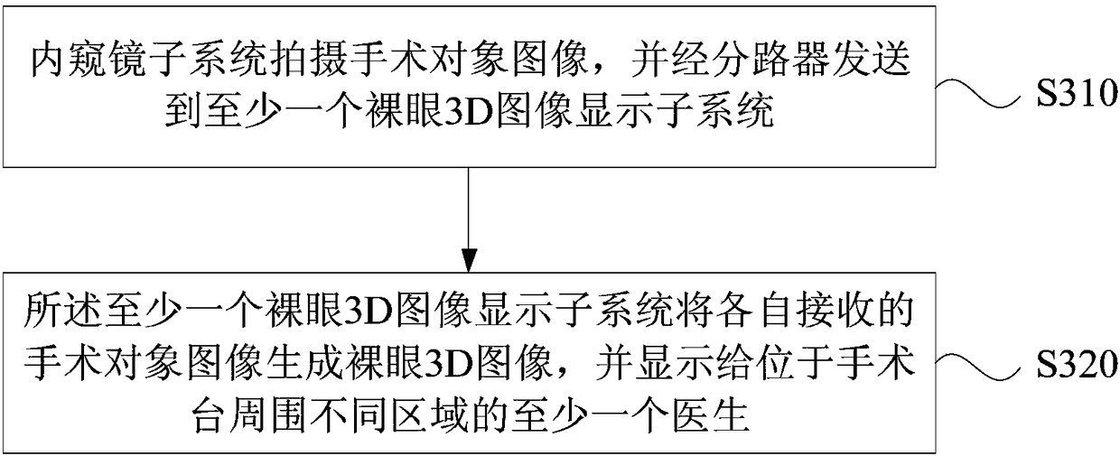
[0039] image 3 The flow chart of the method for displaying a naked-eye 3D image in endoscopic surgery provided by Embodiment 2 of the present invention can be realized by the system for displaying a naked-eye 3D image in endoscopic surgery in the embodiment of the present invention. The method specifically includes:

[0040] S310. The endoscope system captures images of the surgical object, and sends them to at least one naked-eye 3D image display subsystem through a splitter.

[0041]Specifically, an endoscope is a tube equipped with a light that can be passed through the mouth into the stomach or into the body through other natural orifices. The endoscope can be used to see lesions that cannot be seen on X-rays, so it is very useful for doctors. For example, with the help of an endoscope, doctors can observe ulcers or tumors in the stomach, and formulate the best treatment plan accordingly, or use the endoscope to perform minimally invasive surgery. After the endoscope e...

Embodiment 3

[0050] Figure 4 It is a flow chart of the naked-eye 3D image display method for endoscopic surgery provided by Embodiment 3 of the present invention, which is further optimized on the basis of the above embodiments, and illustrates how to perform face recognition and obtain image interleaving parameters. Such as Figure 4 As shown, the naked-eye 3D image display method for endoscopic surgery includes the following steps:

[0051] S410. The endoscope system captures images of the surgical object, and sends them to at least one naked-eye 3D image display subsystem through a splitter.

[0052] S420. Each dual-camera eye tracking device in the at least one naked-eye 3D image display subsystem captures the doctor's facial image, determines the spatial position of the human eye according to the facial image, and determines the image interleaving parameter according to the spatial position of the human eye.

[0053] The dual-camera eye tracking device captures and recognizes the d...


PUM


Abstract

An embodiment of the invention discloses an endoscopic surgery glasses-free 3D image display system and method, wherein the system comprises an endoscopic subsystem, a shunt and at least one glasses-free 3D image display subsystem; the endoscopic subsystem is connected with all glasses-free 3D image display subsystems through the shunt and is used for taking an image of a surgical subject; the image of the surgical subject is transmitted to each glasses-free 3D image display subsystem through the shunt; the at least one glasses-free 3D image display subsystem is used for generating a glasses-free 3D image via the received images of the surgical subject, and displaying the glasses-free 3D image to at least one surgeon in different areas around an operating table. The problems are solved that glasses devices need to be put on when traditional polarizing 3D systems are in use, and multiple surgeons engaging in surgery cannot view at different angles; therefore, surgical burden of the surgeons is relieved; multiple surgeons can view the 3D image of a surgical subject, so that surgical safety is improved.

Description

technical field [0001] Embodiments of the present invention relate to the technical field of medical equipment, and in particular to a naked-eye 3D image display system and display method for endoscopic surgery. Background technique [0002] With the development of 3D image display technology, 3D display has gradually replaced the application of 2D display technology in the field of laparoscopic surgery. The application of 3D image display technology in laparoscopic surgery can improve the accuracy of identification of the internal structure of the human body, reduce the learning curve of minimally invasive surgery for doctors, and enhance the safety of surgery. [0003] Generally, the 3D image is captured by a dual-camera endoscope, and its display is completed by a traditional polarized 3D display. However, when using the traditional polarized 3D system, all users need to wear glasses. Due to the long operation time, it will bring additional burden to doctors, especially ...

Claims


Application Information

Patent Timeline
Patent Type & Authority Applications(China)
IPC IPC(8): A61B1/04
CPCA61B1/00193A61B1/04
Inventor 谢春华高金城于炀陈佳搏潘燕峰潘熙松李国新胡彦锋
Owner ZHANGJIAGANG KANGDE XIN OPTRONICS MATERIAL
Why Eureka
Try Eureka