Method for automatically segmenting liver tumors in abdominal CT sequence images

A technology for sequential images and liver tumors, applied in the field of image processing, can solve problems such as complex structures, blurred borders of liver tumors, and low contrast

Active Publication Date: 2018-09-28
HUNAN UNIV OF SCI & TECH
View PDF3 Cites 21 Cited by
  • Summary
  • Abstract
  • Description
  • Claims
  • Application Information

AI Technical Summary

Problems solved by technology

[0004] The purpose of the present invention is to provide a method for automatic segmentation of liver tumors in abdominal CT sequence images, aiming to solve the problem of automatic segmentation of liver tumors cause

Method used


Image

  • Method for automatically segmenting liver tumors in abdominal CT sequence images
  • Method for automatically segmenting liver tumors in abdominal CT sequence images
  • Method for automatically segmenting liver tumors in abdominal CT sequence images

Examples


Embodiment 1

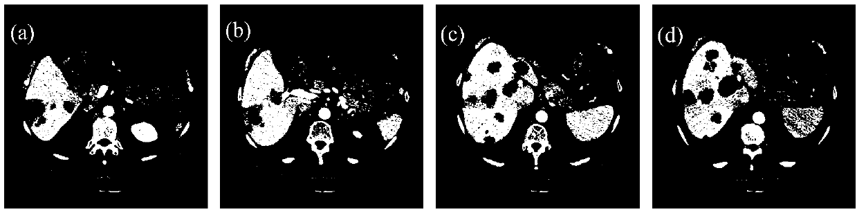
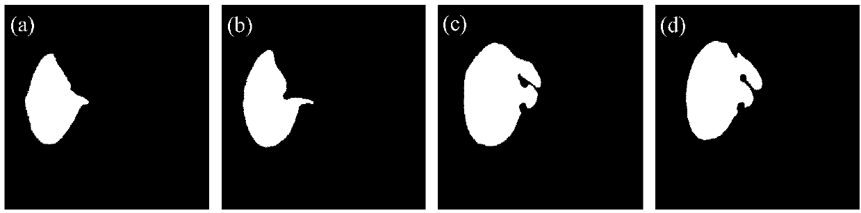
[0037] In order to obtain the liver region in the abdominal CT sequence images, the automatic abdominal CT sequence liver segmentation method published in the document "A hierarchical local region-based sparse shape composition for liver segmentation in CT scans" (pattern recognition, pp.88-106, 2016.) was used. The original CT sequence images are preprocessed to obtain the liver region in the sequence. figure 1 (a)-(d) are 4 original images randomly selected from a certain CT sequence, figure 2 (a)-(d) are the liver segmentation results obtained by using the method of this embodiment, that is, the liver region mask.

Embodiment 2

[0039] A liver area enhancement method for abdominal CT sequence images, the specific implementation steps are as follows:

[0040] (1) Using Embodiment 1 to obtain the liver region in the abdominal CT sequence image f;

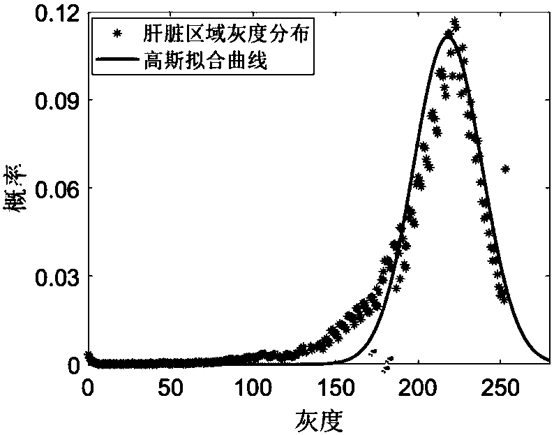
[0041] (2) In order to obtain the gray distribution range of the liver region, a Gaussian function is used to fit the gray histogram of the entire liver region in the sequence:

[0042]

[0043] Among them, c is the peak value of the Gaussian distribution, and μ and σ are the center and standard deviation of the Gaussian distribution, respectively. image 3 for right figure 1 The Gaussian fitting result of the gray histogram of the liver area in the sequence image shown, it can be seen that the gray level of the liver can better conform to the Gaussian distribution. According to the probability theory of Gaussian distribution, the gray ranges of [μ-σ,μ+σ], [μ-2σ,μ+2σ] and [μ-3σ,μ+3σ] occupy 68%, 95%, 99% of the pixels. Considering the noise and tumor t...

Embodiment 3

[0050] Using Example 2 to obtain liver enhancement results, combined with image boundary information, construct a graph cut energy function for multi-target segmentation:

[0051]

[0052] Among them, α is a normal number from 0 to 1, P represents all pixel sets in the abdominal CT sequence image f, N p Represents the neighborhood pixel set of pixel p, R(f p ) and B(f p , f q ) are the grayscale and boundary penalty items, and are obtained by the following formulas:

[0053]

[0054]

[0055] in

[0056]

[0057]

[0058] f p and f q respectively represent the pixel points p and q in the image f, I p and I q Represents the gray value of pixels p and q, d(p,q) represents the Euclidean distance between pixels p and q, T P Indicates the total number of pixels in the pixel set P of the image f, mask is the liver mask obtained in Example 1, the pixels belonging to the liver area are marked as 1, and the pixels belonging to the background are marked as 0, that...


PUM


Abstract

The invention discloses a method for automatically segmenting liver tumors in abdominal CT sequence images. The method comprises the following steps of: preprocessing: preprocessing an abdominal CT sequence image so as to obtain a liver area in the image; liver enhancement: improving a contrast ratio of normal liver parenchyma to tumor tissue by adoption of a segmented nonlinear enhancement operation and an iterative convolution operation according to a grey level distribution characteristic of the liver area; automatic segmentation: constructing an image segmentation energy function for multi-target segmentation by utilizing the enhancement result and combining image boundary information, minimizing the energy function by adoption of an optimal algorithm and obtaining a liver tumor preliminary automatic segmentation result; and post-processing: optimizing the preliminary segmentation result by adoption of a three-dimensional mathematic morphological open operation, and removing mis-segmented areas to improve the segmentation precision. The method is beneficial for helping radiological experts and surgeons to timely and effectively obtaining overall information and three-dimensional display of liver tumors and providing technical support for computer-aided diagnosis and treatment of liver diseases.

Description

technical field [0001] The invention belongs to the technical field of image processing and relates to target segmentation in medical images, in particular to automatic segmentation of liver tumor tissue in abdominal CT sequence images, which can be used for medical image auxiliary diagnosis and treatment. Background technique [0002] More than 50% of new and dead liver cancer patients in the world occur in China, and about 300,000 people die of liver cancer in China every year. Since the symptoms of early liver cancer are not obvious, about 60% of patients do not go to see a doctor until they feel unwell. At this time, they often enter the middle and late stages and lose the chance of radical treatment. Statistics show that the survival rate of patients with advanced liver cancer within 5 years is only about 7%. [0003] Liver tumor burden analysis is usually used to monitor the disease evolution of liver cancer patients, formulate treatment plans, compare different treat...

Claims


Application Information

Patent Timeline
IPC IPC(8): G06T7/00G06T7/11G06T7/194G06T5/00G06T5/30G06T5/40
CPCG06T5/002G06T5/30G06T5/40G06T7/0012G06T7/11G06T7/194G06T2207/10081G06T2207/30056G06T2207/30096
Inventor 廖苗赵于前刘毅志方志雄欧阳军林
Owner HUNAN UNIV OF SCI & TECH
Why Eureka
Try Eureka