X-ray image feature detection and registration systems and methods

An x-ray, registration technique used in the detection and analysis of image features

Active Publication Date: 2018-10-09
LIGHTLAB IMAGING
View PDF5 Cites 3 Cited by
  • Summary
  • Abstract
  • Description
  • Claims
  • Application Information

AI Technical Summary

Problems solved by technology

Therefore, all of these factors and others make it challenging to perform diagnostic intravascular imaging and registration as part of the image display and diagnostic process
In addition, challenges related to imaging, image analysis, and registration can negatively impact stent planning and other procedures that rely on these imaging data and associated diagnostic information

Method used


Image

  • X-ray image feature detection and registration systems and methods
  • X-ray image feature detection and registration systems and methods
  • X-ray image feature detection and registration systems and methods

Examples


Embodiment Construction

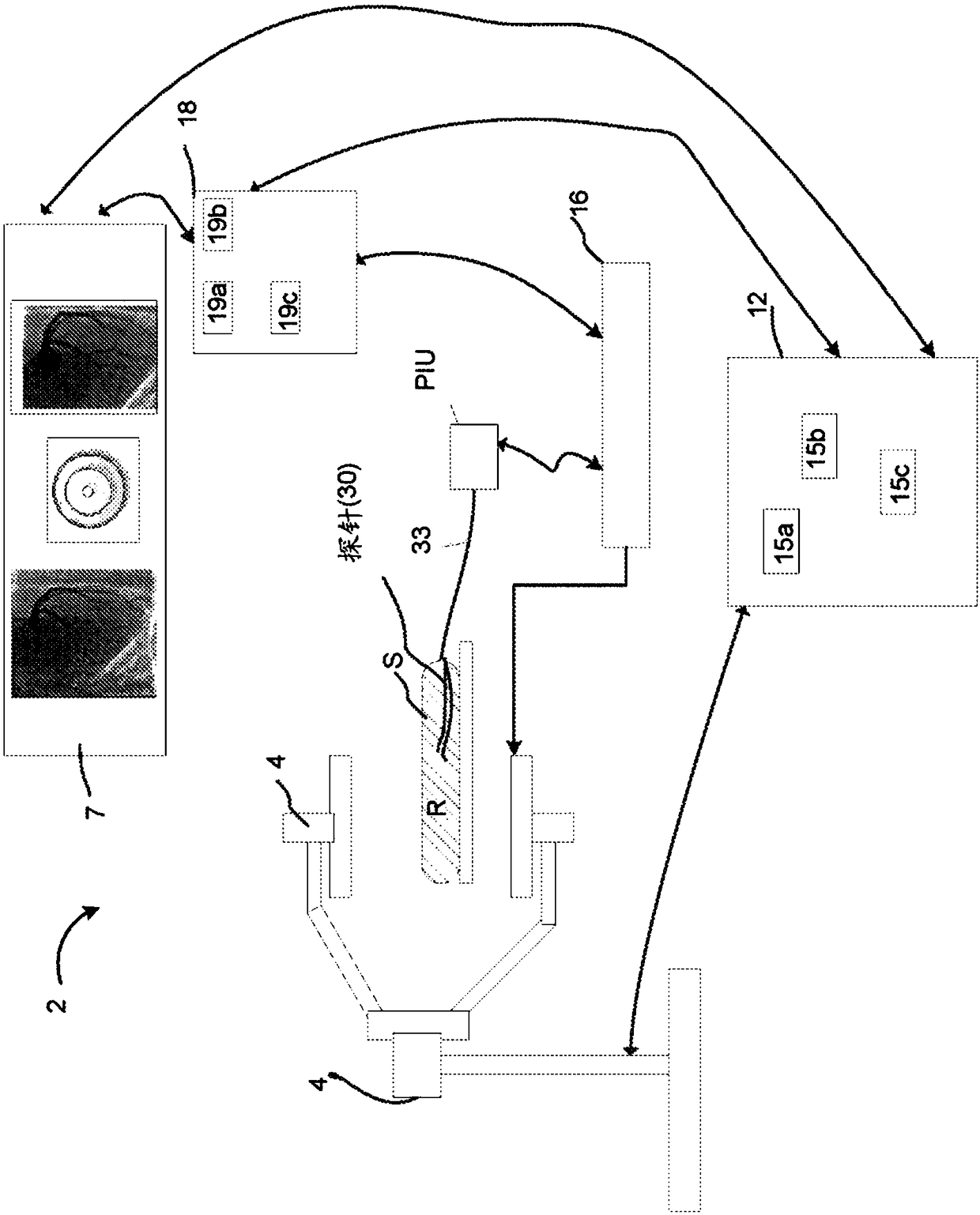
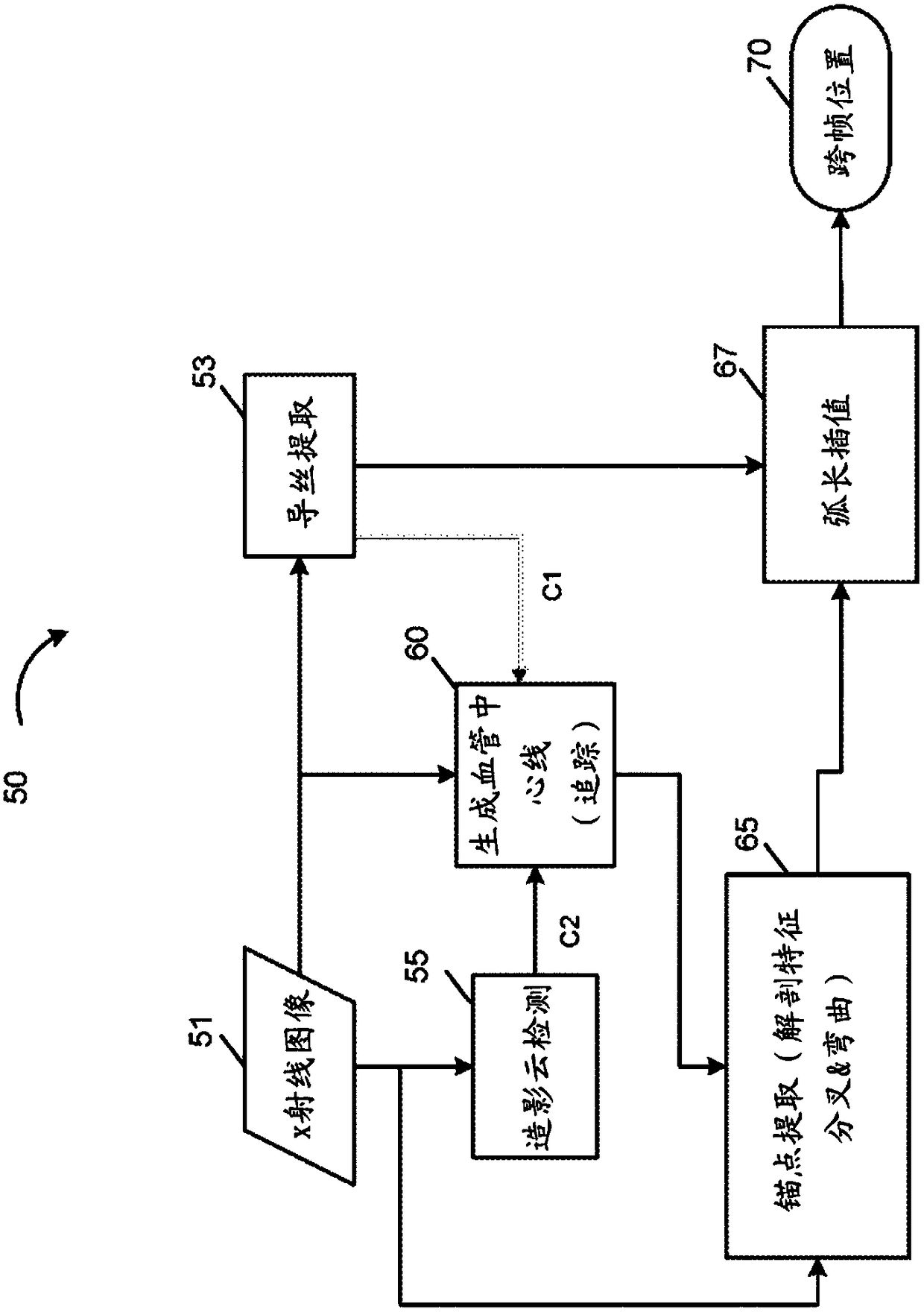
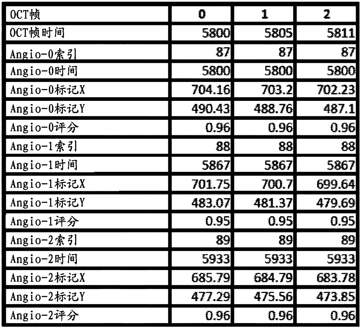
[0072]The present invention relates to various methods, systems and instruments related to x-ray imaging, such as angiography and its use in cardiology. In particular, the invention relates to features related to the co-registration of frames of angiography data across or between these frames. The invention also relates to various methods of improving such co-registration, such as by reducing errors or detecting structures associated with frames of angiography data.

[0073] As examples of this error reduction approach and other angiographic or peripheral vasculature imaging enhancements, several examples are discussed in detail herein. These embodiments relate to contrast cloud detection, extraction or identification of filaments in a frame of x-ray image data, and tracking or registration of features and devices relative to the vasculature, including with respect to angled branches and bifurcations or guide wires. These implementations reduce errors that can propagate throu...


PUM


Abstract

The invention relates generally to the field of vascular system and peripheral vascular system data collection, imaging, image processing and feature detection relating thereto. In part, the disclosure more specifically relates to methods for detecting position and size of contrast cloud in an x-ray image including with respect to a sequence of x-ray images during intravascular imaging. Methods ofdetecting and extracting metallic wires from x-ray images are also described herein such as guidewires used in coronary procedures. Further, methods for of registering vascular trees for one or moreimages, such as in sequences of x-ray images, are disclosed. In part, the disclosure relates to processing, tracking and registering angiography images and elements in such images. The registration can be performed relative to images from an intravascular imaging modality such as, for example, optical coherence tomography (OCT) or intravascular ultrasound (IVUS).

Description

[0001] Cross References to Related Applications [0002] This application claims priority and benefit to US Provisional Patent Application no. 62 / 257,213, filed November 18, 2015, the disclosure of which is incorporated herein by reference in its entirety. technical field [0003] In part, the present invention relates generally to the field of vasculature and peripheral vasculature imaging and data acquisition. More specifically, and in part, the present invention relates to the detection and analysis of image features. Background technique [0004] X-ray images provide an important diagnostic tool in various disciplines. For example, interventional cardiologists use x-ray-based imaging systems in a variety of procedures and exams. As a specific type of x-ray imaging, angiographic imaging of blood vessels is often performed using fluoroscopy. Visualization of arteries during catheterization is a valuable diagnostic tool. This visualization aids in the planning and exec...

Claims


Application Information

Patent Timeline
IPC IPC(8): G06T5/50
CPCG06T5/50G06T2207/10116G06T2207/30101G06T7/33A61B6/5247A61B6/504G06T7/0012A61B5/0066A61B8/12G06T2207/10101G06T7/73G06T7/0014G06T2207/10016G06T2207/10132G06T2207/30021G06T2207/30104G06T2207/30204
Inventor 罗琳娜·达斯卡尔伊泰·温克勒
Owner LIGHTLAB IMAGING
Why Eureka
Try Eureka