Eureka AIR delivers breakthrough ideas for toughest innovation challenges, trusted by R&D personnel around the world.

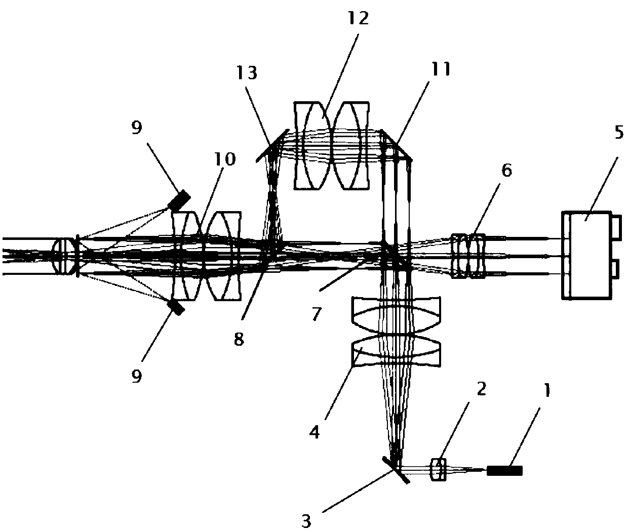
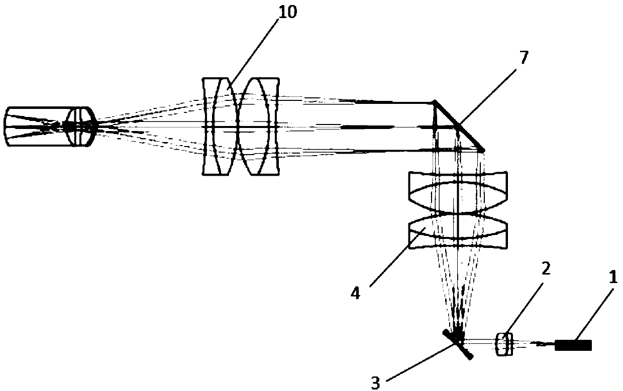
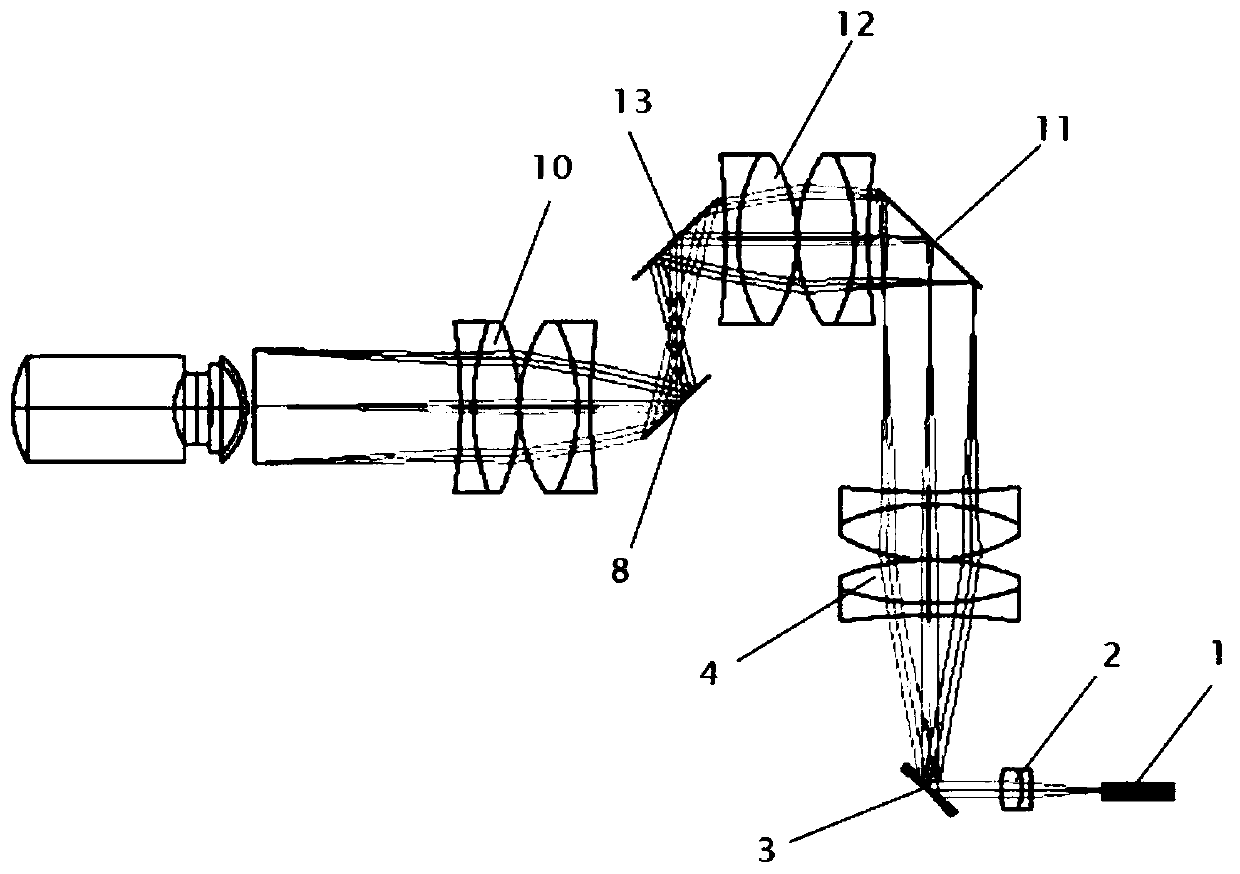
Optical path structure of two-channel OCT (optical coherence tomography) sample arm in posterior segment and anterior segment of eye

An optical coherence tomography and sample arm technology, which is used in medical imaging, medical science, and eye testing equipment, etc. Effect

Pending Publication Date: 2018-11-06
江苏伊士嘉医疗科技有限公司
View PDF4 Cites 3 Cited by
  • Summary
  • Abstract
  • Description
  • Claims
  • Application Information

AI Technical Summary

Problems solved by technology

However, in order to maintain the miniaturization of the handheld probe, the iris camera used in this system for auxiliary system and human eye alignment can only be used in the anterior segment OCT imaging mode
However, considering the limitation of the pupil on the light beam, the alignment of the optical path in the process of OCT imaging of the posterior segment of the eye is more difficult, and the assistance of the iris camera is more needed

Method used


Image

  • Optical path structure of two-channel OCT (optical coherence tomography) sample arm in posterior segment and anterior segment of eye
  • Optical path structure of two-channel OCT (optical coherence tomography) sample arm in posterior segment and anterior segment of eye
  • Optical path structure of two-channel OCT (optical coherence tomography) sample arm in posterior segment and anterior segment of eye

Examples


Embodiment Construction

[0027] The following will clearly and completely describe the technical solutions in the embodiments of the present invention with reference to the accompanying drawings in the embodiments of the present invention. Obviously, the described embodiments are only some, not all, embodiments of the present invention. Based on the embodiments of the present invention, all other embodiments obtained by persons of ordinary skill in the art without making creative efforts belong to the protection scope of the present invention.

[0028] see Figure 1~4, in an embodiment of the present invention, a dual-channel optical coherence tomography sample arm optical path structure of anterior segment and posterior segment includes an optical fiber connector 1, an optical fiber collimator 2, a scanning galvanometer 3, a first lens 4, an iris camera 5, OCT sample of the anterior segment formed by the fourth lens 6, the first dichroic mirror 7, the second dichroic mirror 8, LED9, the third lens 10...


PUM


Abstract

The invention discloses an optical path structure of a two-channel OCT (optical coherence tomography) sample arm in a posterior segment and an anterior segment of an eye. The optical path structure comprises three optical paths: an anterior segment OCT sample arm optical path, a posterior segment OCT sample arm optical path and an iris imaging optical path. According to the structure, imaging modes of the anterior segment and the posterior segment are switched through the rotation of a dichroic mirror, thereby avoiding the secondary attenuation of the signal intensity by a conventional beam splitter, and each OCT imaging mode can be assisted to be calibrated by the LED illuminated iris imaging optical path.

Description

technical field [0001] The invention relates to the technical field of medical imaging equipment, in particular to a dual-channel optical coherence tomography sample arm optical path structure for anterior segment and posterior segment. Background technique [0002] Optical coherence tomography (OCT) is a high-resolution, high-speed, non-destructive optical imaging technology invented by researchers at MIT in the 1990s, which can provide three-dimensional structural imaging for biological tissues. Since its invention, it has been widely used in biomedical research and clinical aspects, such as histomorphological research and real-time navigation of ophthalmic clinical operations. As a unique application of the optical coherence tomography system, OCT technology has very important application value and great application prospect in the diagnosis, screening and intraoperative navigation evaluation of ophthalmic diseases. [0003] Although the earliest and most widely used app...

Claims


Application Information

Patent Timeline
Patent Type & Authority Applications(China)
IPC IPC(8): A61B3/10A61B3/14
CPCA61B3/0008A61B3/102A61B3/14A61B2576/02
Inventor 黄勇杨爱岭张丰尧张松
Owner 江苏伊士嘉医疗科技有限公司
Why Eureka
Eureka Blog
Learn More