Blood cell component analysis instrument and method

A component analysis and blood cell technology, applied in the field of medical inspection, can solve problems such as high failure rate, inability to accurately identify cells, and low work efficiency

Inactive Publication Date: 2018-11-20
CLINDIAG SYSTEMS CO LTD
View PDF4 Cites 11 Cited by
  • Summary
  • Abstract
  • Description
  • Claims
  • Application Information

AI Technical Summary

Problems solved by technology

The electrical impedance method blood analyzer has the disadvantages of high reagent consumption, high failure rate, and heavy instrument maintenance workload.
The detection accuracy of flow cytometry has been improved, but the instrument is complicated and the cost of reagents is extremely high
The traditional manual microscope method has extremely high requirements on the operator's skills and eyesight, the work intensity is extremely high, and the work efficiency is extremely low
Although the dry blood analyzer consumes less reagents and has a lower failure rate, the detection information is simple and the cells cannot be accurately identified

Method used


Image

  • Blood cell component analysis instrument and method
  • Blood cell component analysis instrument and method
  • Blood cell component analysis instrument and method

Examples


Embodiment 1

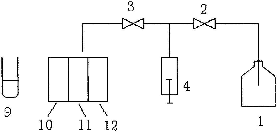
[0047] Such as figure 1 , in this embodiment, the working principle of the sample suction and transfer unit: put the special detection card on the detection card position of the blood cell component analyzer, close the first valve 2, open the second valve 3, pull down the first syringe 4, and the instrument The sample aspirating needle draws an equal amount of sample from the sample tube 9 into the syringe 4, then the aspirating needle is transferred to the top of the test card, the first syringe 4 is pushed up, and the equal amount of sample is transferred to the control tube 10, the first detection tube 11 and the control tube 10. In the second detection tube 12, then turn on the mixing motor under the detection card position, and mix the blood samples in the control tube and the detection tube on the detection card respectively;

[0048] The cleaning needle is transferred to the top of the cleaning pool, the first valve 2 is opened, the second valve 3 is closed, the syringe...

Embodiment 2

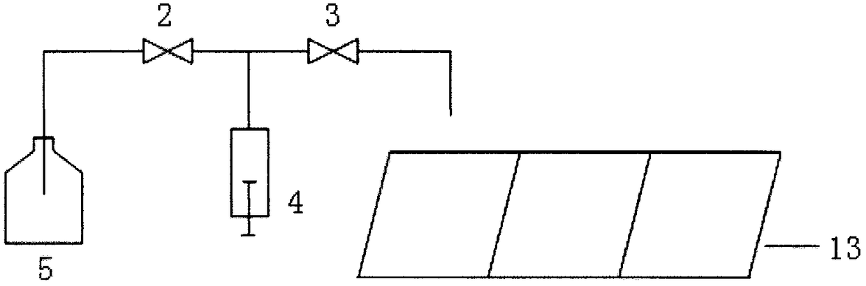
[0051] Such as Figure 4 , In this embodiment, the stirring and smearing unit is an inverted T-shaped stirring and smearing unit. When the blood sample and staining solution are added to the slide glass, the inverted T-shaped stirring and smear unit 6 moves vertically above the slide glass, and its cross arm descends to a position greater than 0.1mm from the surface of the slide glass, and the inverted T-shaped stirring and The smear unit rotates to mix the mixture 8 of the blood sample and the staining solution; then the inverted T-shaped stirring and the smear unit continue to descend and rotate close to the glass slide, and then the inverted T-shaped stirring and the smear unit rises and shifts, Cling to the glass slide again and rotate it to spread the blood sample on another smear area of ​​the glass slide, and then move the T-shape stirring and smearing unit to the cleaning tank for cleaning.

Embodiment 3

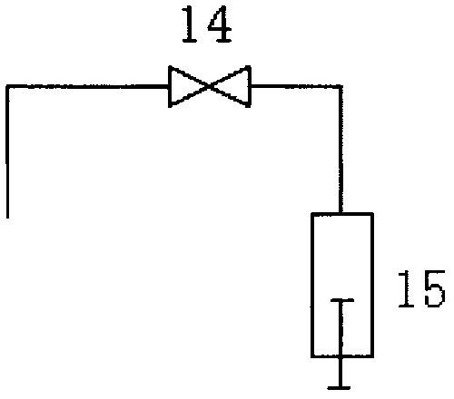
[0053] Such as image 3 , in this embodiment, the instrument is not provided with a cleaning solution bottle, but is provided with a cleaning pool. When the needle is cleaned, the sampling needle is transferred to the cleaning pool, the third valve 14 is opened, the second syringe 15 is pulled down, and the sampling needle is removed from the cleaning pool. The cleaning water is sucked into the second syringe, and then the sampling needle is transferred to the top of the waste liquid collector, the second syringe is pushed up, and the cleaning water is discharged to the waste liquid collector.


PUM


Abstract

The invention provides a blood cell component analysis instrument and a method. The instrument comprises a sample absorbing and transfer unit, a dyeing liquid adding unit, a stirring and smearing unit, a cleaning unit, a microscanning unit, an image processing unit and a control unit. The sample absorbing and transfer unit is used for absorbing a quantitative blood sample to a glass slide; the dyeing liquid adding unit is used for adding the dyeing liquid to dye cells and dilute the blood sample; a reverse T-shaped stirring and smearing unit is used for mixing the blood sample and the dyeing liquid uniformly and smearing the mixed blood sample; the microscanning unit is used for performing microscopic examination on the smear; the instrument automatically and continuously performs axial scanning on the unclear targets for many times; the image processing unit processes the scanning image and selecting the images with clearest cells or granules in the multi-focus images into a whole image; and the analysis instrument is simple to operate, low in failure rate, accurate in result and few in consumed material reagent, and can perform quantitative analysis on the blood cells conveniently and accurately.

Description

technical field [0001] The invention relates to the field of medical testing, in particular to a blood cell component analyzer and method. Background technique [0002] The working principles of existing hematology analyzers are mainly electrical impedance method, flow cytometer, manual microscope inspection and dry centrifugal scanning method. The resistance method counts the number of particles passing through the micropores of the instrument and the size of the particles for classification and counting. It cannot distinguish the shape and internal structure of blood cells, so the classification of cells is not accurate enough; and each cell in blood belongs to a heterogeneous group. The morphology and structure are divided into many different types, and their quantity and function are different. Since the pathological changes of many diseases at the blood cell level are not only reflected in the number of blood cells, the morphological changes of cells also have important...

Claims


Application Information

Patent Timeline
Patent Type & Authority Applications(China)
IPC IPC(8): G01N21/31G01N1/38G01N1/31G01N1/28
CPCG01N1/2813G01N1/31G01N1/38G01N21/314G01N2001/387G01N2021/3129
Inventor 徐新董自权周强詹小勇郭敏李晓兰
Owner CLINDIAG SYSTEMS CO LTD
Why Eureka
Try Eureka