Multi-magnification automatic microscopic imaging method and device

A microscopic imaging and fully automatic technology, applied in the field of optical imaging, can solve the problems of low accuracy of inspection results, heavy workload of inspectors, and low degree of automation, so as to reduce time costs and inspection personnel needs, and reduce inspection costs. , the effect of avoiding motion errors

Inactive Publication Date: 2018-11-20
DIRUI MEDICAL TECH CO LTD
View PDF3 Cites 4 Cited by
  • Summary
  • Abstract
  • Description
  • Claims
  • Application Information

AI Technical Summary

Problems solved by technology

[0003] Based on this, it is necessary to provide a multi-magnification fully automatic microscopic device that is currently used in clinical morphological testing due to the low degree of automation, resulting in low accuracy of test results, heavy workload of test personnel, and low test efficiency. Microscopic imaging method and device

Method used


Image

  • Multi-magnification automatic microscopic imaging method and device
  • Multi-magnification automatic microscopic imaging method and device
  • Multi-magnification automatic microscopic imaging method and device

Examples


Embodiment Construction

[0022] The technical solutions of the present invention will be described in detail below in conjunction with the accompanying drawings and preferred embodiments.

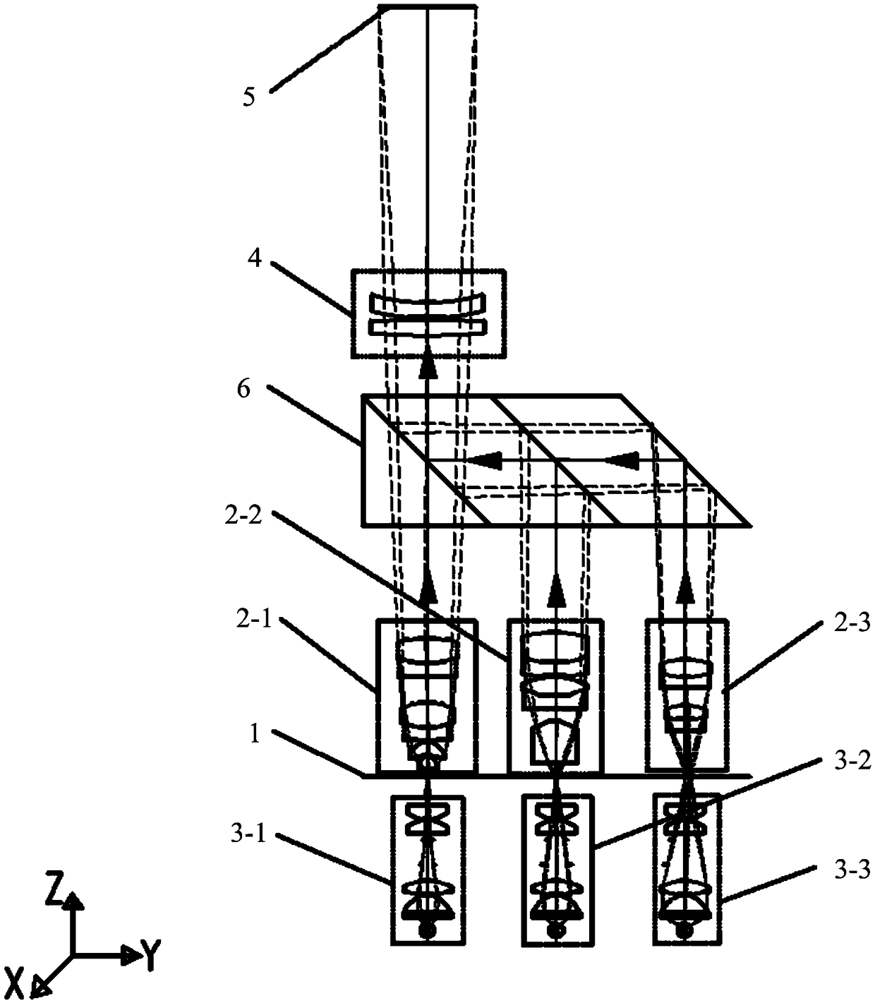
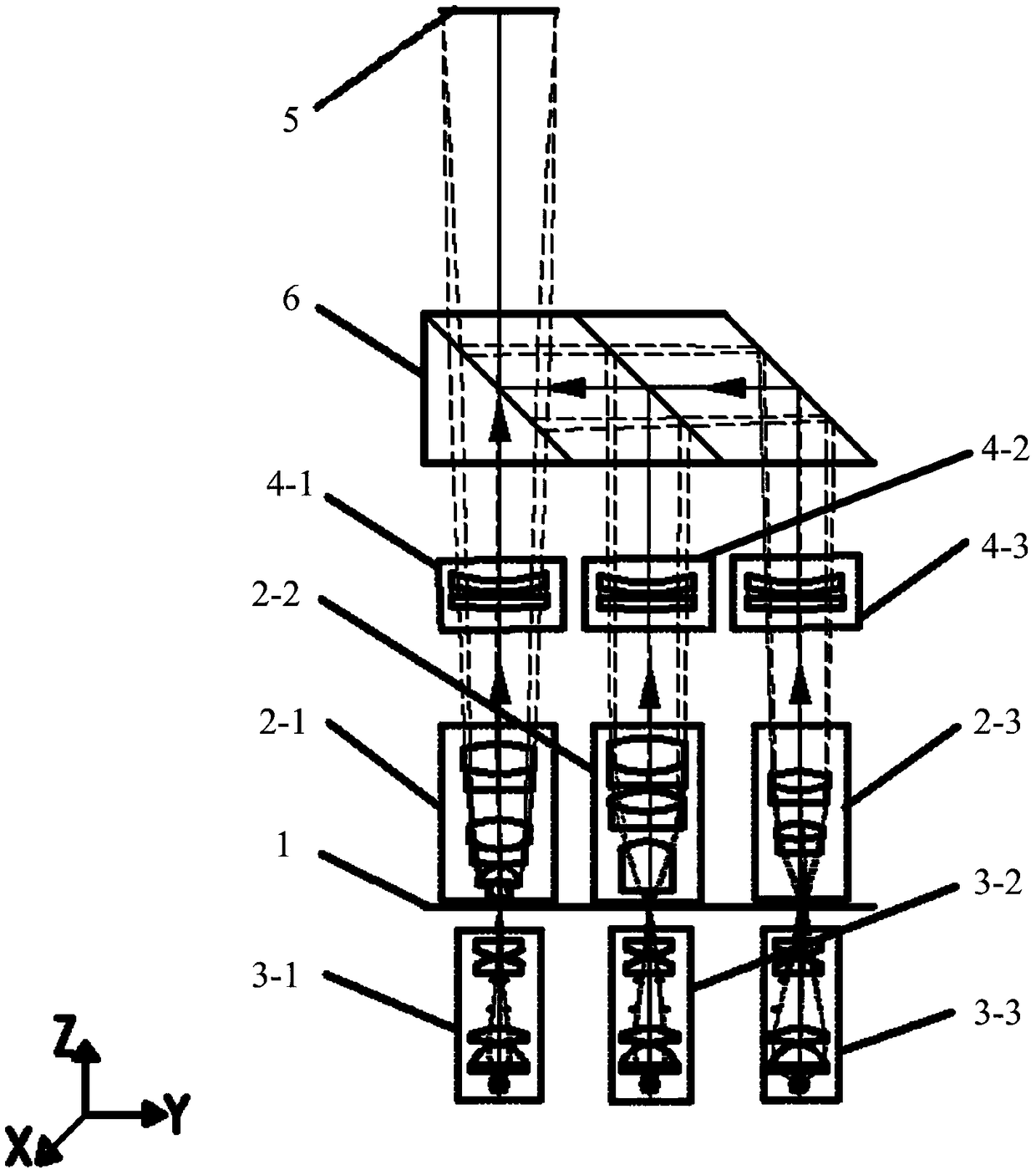
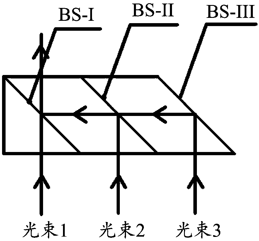
[0023] In one of the examples, as Figure 1-Figure 3 As shown, the multi-magnification automatic microscopic imaging device includes a three-dimensional motion platform 1, at least two microscopic objective lenses, an illumination unit, a tube lens 4, an imaging unit 5, a beam integration component 6 and a fully automatic control unit.

[0024] Specifically, the three-dimensional motion platform 1 can also be called a sample stage, which is used to carry the sample of the measured object. The three-dimensional motion platform 1 can perform three-dimensional independent movement under the control of a fully automatic control unit, and is parallel to the direction of the optical axis of the microscope (that is, the Z direction). ) has the highest motion accuracy, which can reach the micron level or even higher.

[0...


PUM


Abstract

The invention relates to a multi-magnification automatic microscopic imaging method and device and belongs to the technical field of optical imaging. The device comprises a three-dimensional motion platform used for bearing a detected sample, at least two microscope objectives with different magnifications, an illumination unit corresponding to the microscope objectives, a tube mirror arranged above the microscope objectives, a beam integration component arranged between the microscope objectives and the tube mirror or between the tube mirror and an imaging unit, the imaging unit used for imaging the detected sample, and an automatic control unit connecting with the three-dimensional motion platform and the imaging unit, wherein the automatic control unit is used for controlling three-dimensional movement of the three-dimensional motion platform and brightness of the illumination unit, and an intelligible image containing a detected target object is obtained. The device is advantaged in that the need for objective or tube mirror switching motion when different magnification requirements are for imaging is avoided, so the motion error caused by the motion structure is avoided, and the microscopic imaging quality and detection efficiency are improved.

Description

technical field [0001] The invention relates to the technical field of optical imaging, in particular to a multi-magnification automatic microscopic imaging method and device. Background technique [0002] In the process of continuous development of the optical microscope, its function is becoming more and more powerful. In the microscopic imaging device, in order to meet the morphological microscopic imaging detection requirements of the measured object, the microscopic imaging device needs to be equipped with microscopic objectives with different magnifications. . In the actual use of the microscope, manual operations are mostly used, that is, it is necessary to manually place the measured object on the stage, rotate and replace the objective lens to be used, manually focus, adjust brightness, find the measured object, morphological analysis and judgment and For operation steps such as classification and counting, the operator not only has a heavy workload, takes a long t...

Claims


Application Information

Patent Timeline
Patent Type & Authority Applications(China)
IPC IPC(8): G02B21/00G02B21/06G02B21/36
CPCG02B21/0032G02B21/0052G02B21/06G02B21/361
Inventor 石欣陈晨沈晓晨
Owner DIRUI MEDICAL TECH CO LTD
Why Eureka
Try Eureka