System and method for ear-nose-throat examination image screening control, and application

A control method, ENT technology, applied in the medical field, can solve problems that affect the detection effect and cannot be obtained

Inactive Publication Date: 2018-11-27
THE THIRD XIANGYA HOSPITAL OF CENT SOUTH UNIV
View PDF11 Cites 1 Cited by
  • Summary
  • Abstract
  • Description
  • Claims
  • Application Information

AI Technical Summary

Problems solved by technology

It is impossible to find all the solutions; how to find all the solutions for the absolute va

Method used


Image

  • System and method for ear-nose-throat examination image screening control, and application
  • System and method for ear-nose-throat examination image screening control, and application
  • System and method for ear-nose-throat examination image screening control, and application

Examples


Embodiment Construction

[0068] In order to further understand the content, features and effects of the present invention, the following examples are given, and detailed descriptions are given below with reference to the accompanying drawings.

[0069] The structure of the present invention will be described in detail below in conjunction with the accompanying drawings.

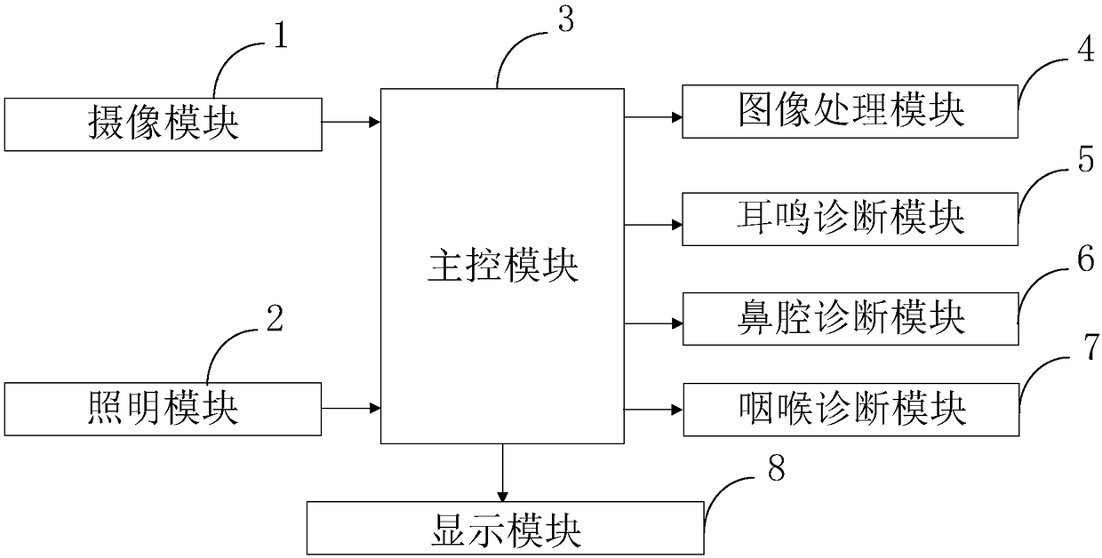
[0070] Such as figure 1 As shown, the ENT inspection image screening control system provided by the present invention includes: camera module 1, lighting module 2, main control module 3, image processing module 4, tinnitus diagnosis module 5, nasal cavity diagnosis module 6, throat diagnosis module 7, Module 8 is displayed.

[0071] The camera module 1 is connected with the main control module 3, and is used to insert and collect image data on the patient's ear, nasal cavity and throat through a miniature camera;

[0072] The lighting module 2 is connected with the main control module 3, and is used to provide the lighting function...


PUM


Abstract

The invention belongs to the technical field of medical treatment, and discloses a system and a method for ear-nose-throat examination image screening control, and an application. The control system comprises: a camera shooting module, a lighting module, a main control module, an image processing module, a tinnitus diagnosis module, a nasal cavity diagnosis module, a throat diagnosis module, and adisplay module. The method include: collecting noise images of examination position images of an ear, a nasal cavity, and a throat by using a basic model of a fully differential reconstruction algorithm based on a non-local mean regularization item through the camera shooting module; through the image processing module, processing the images collected by the camera shooting module; using a pure tone threshold detection method to detect pure tone hearing thresholds of both ears through the tinnitus diagnosis module. The system and the method greatly improve reliability of examined image and are beneficial to a doctor for accurate diagnosis. At the same time, the tinnitus diagnosis module greatly shortens matching time of tinnitus frequency, increases fault tolerance capability, and solvesa problem in the prior art that tinnitus frequency is possible to be leaked in detection, and improves diagnosis effect.

Description

technical field [0001] The invention belongs to the field of medical technology, and in particular relates to an ENT inspection image screening control system, method and application. Background technique [0002] At present, the existing technologies commonly used in the industry are as follows: [0003] Otolaryngology examination must be cautious, because otolaryngology is a deep small cavity, so special lighting devices and examination instruments must be used for examination. Inflatable otoscope, gun-like tweezers, cotton roll, cerumen hook, tongue depressor, anterior rhinoscope, posterior rhinoscope, indirect laryngoscope, tuning fork, sprayer, etc. However, the existing ENT examination images are unclear and unreliable, which affects the doctor's diagnosis; at the same time, the existing diagnosis efficiency for tinnitus is low, and the frequency of tinnitus is easily missed, resulting in poor diagnostic effect. [0004] In summary, the problems in the prior art are:...

Claims


Application Information

Patent Timeline
IPC IPC(8): G06T7/00G06T5/00
CPCG06T5/002G06T7/0012
Inventor 宋业勋李维张永全李和清刘火旺张晓伟
Owner THE THIRD XIANGYA HOSPITAL OF CENT SOUTH UNIV
Why Eureka
Try Eureka