Anti-human PD1 monoclonal antibody and application thereof

A monoclonal antibody, antibody technology, applied in the direction of antibody, anti-animal/human immunoglobulin, application, etc., can solve the problems of lack of MYPPY, inability to bind B7-1 and B7-2, etc., to achieve the effect of novel sequence

Inactive Publication Date: 2018-12-07
BIOSION INC
View PDF3 Cites 4 Cited by
  • Summary
  • Abstract
  • Description
  • Claims
  • Application Information

AI Technical Summary

Problems solved by technology

[0004] Although PD1 is structurally similar to CTLA4, it

Method used


Image

  • Anti-human PD1 monoclonal antibody and application thereof
  • Anti-human PD1 monoclonal antibody and application thereof
  • Anti-human PD1 monoclonal antibody and application thereof

Examples


Example Embodiment

[0039] Example 1 Obtaining specific anti-PD1 mouse monoclonal antibodies by fusion hybridoma technology

[0040] 1.1 Animal immunity

[0041] The mice were immunized according to the general method in the literature (E Harlow, D. Lane, Antibody: A Laboratory Manual, Cold Spring Harbor Laboratory Press, Cold Spring Harbor, N.Y., 1998). The immunogen is a recombinant human PD1 (amino acid range Leu 25-Gln 167) protein (Acrobiosystems, Cat#PD1-H5257) with a human IgG1Fc tag at the C-terminus. The recombinant human PD1 protein with his s tag (Sino biological inc., cat#10377-H08H) was used as the detection antigen for serum titer determination and hybridoma screening. Briefly, take out an appropriate amount of Freund's adjuvant into a 1.5ml EP tube and mix it with a shaker. Prepare the antigen protein solution with PBS. Mix the adjuvant and protein antigen solution according to the required amount, push each other through a syringe to fully emulsify the antigen to form a stable water...

Example Embodiment

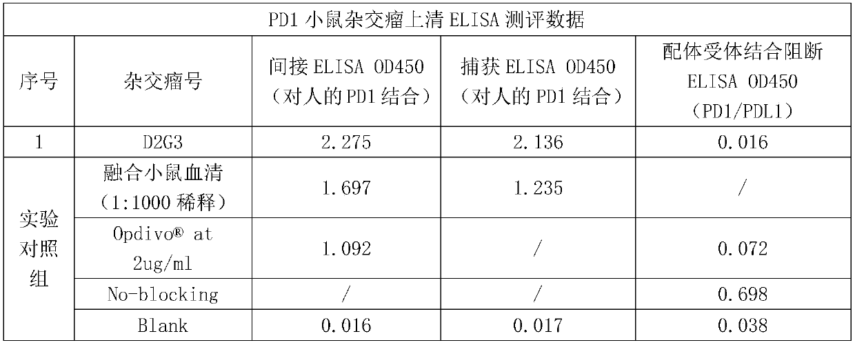
[0047] Example 2 In vitro analysis method for determining the functional activity of PD1 monoclonal antibody

[0048] 2.1 Determination of antibody binding ability based on capture ELSIA

[0049] Prepare goat anti-mouse IgG Fcr-specific secondary antibody (Jackson Immuno Research, #115-006-071) with 1xPBS to make the final concentration 2μg / ml, add 100μl / well to 96-well microplate, 4 degrees Coated overnight. The next day, the plate was washed 4 times with a PBS solution containing 0.05% Tween 20 (ie 1xPBST), and 200 μl / well of 5% skimmed milk powder in PBST solution was added and placed at 37°C for 2 hours. Wash the plate again, add 100μl / well of diluted antibody solution or hybridoma supernatant, incubate at 37°C for 40 minutes, and then wash the plate 4 times. Add the prepared 60nM biotin-labeled human PD1-Fc protein solution (in 2.5% skimmed milk powder in PBST) at 100 μl / well, incubate at 37°C for 40 minutes, and then wash the plate 4 times. Add 1:10000 diluted horseradish ...

Example Embodiment

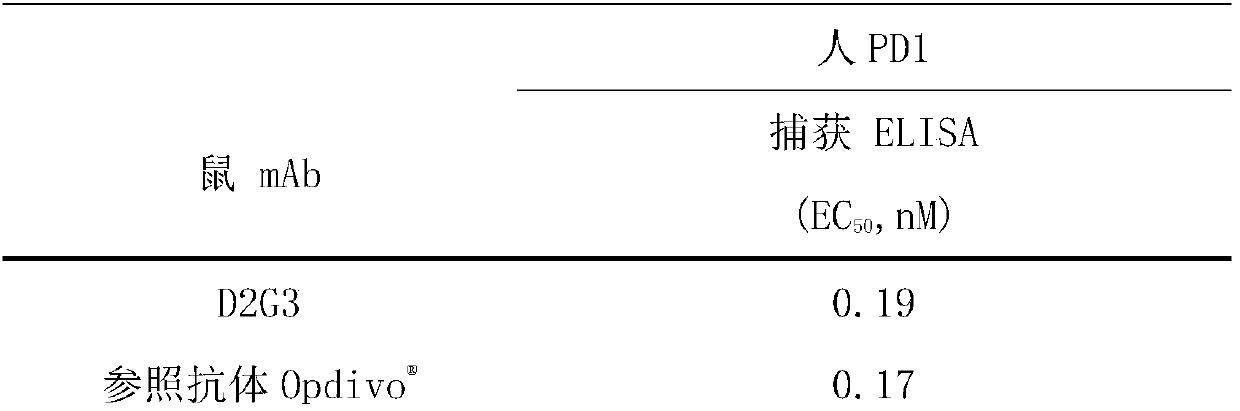
[0059] Example 3 Binding activity of anti-PD1 mouse monoclonal antibody

[0060] According to the analysis method described in Example 2.1, the binding activity of PD1 mouse monoclonal antibody was evaluated and summarized in Table 2 below. Reference antibody As a control, it is an existing commercialized anti-human PD1 monoclonal antibody. It can be seen from Table 2 that the binding activity of the anti-human PD1 monoclonal antibody of the present invention to human PD1 antigen is equivalent to that of the reference antibody.

[0061] Table 2 PD1 antibody binding activity

[0062]

[0063]


PUM


Abstract

The invention relates to an anti-human PD1 monoclonal antibody and application thereof, also provides coding nucleic acid molecules of the antibody, expression carriers, host cells and a method for expressing the antibody, and also provides an antibody immune coupling binder of the antibody, bispecific molecules, chimeric antigen receptors, a pharmaceutical composition and application. Compared with the prior art, the anti-human PD1 monoclonal antibody disclosed by the invention has the following advantages that the monoclonal antibody of human PD1 can be specially identified, and the bindingaffinity of the antibody and the human PD1 is stronger than that of an existing anti-human PD1 monoclonal antibody, and a sequence of the antibody is novel.

Description

technical field [0001] The present invention relates to a high-affinity and functional anti-human PD1 monoclonal antibody or antibody fragment. The present invention also provides the encoding nucleic acid molecules of the antibody, expression vectors, host cells, and methods for expressing the antibody. Also provided are antibody immunoconjugates, bispecific molecules, chimeric antigen receptors, pharmaceutical compositions, and diagnostic and therapeutic methods comprising antibodies of the invention. Background technique [0002] Programmed death receptor 1, also known as PD-1 or CD279, is a member of the CD28 superfamily. PD-1 is mainly expressed on activated B cells, T cells and myeloid cells (Agata et al., supra; Okazaki et al. (2002) Curr.Opin.Immunol.14:391779-82; Bennett et al. (2003 ) J Immunol 170:711-8). Unlike other members of the CD28 family, since there is no unpaired cysteine ​​in the structure of PD-1, it is suggested that PD1 mainly exists in monomeric f...

Claims


Application Information

Patent Timeline
IPC IPC(8): C07K16/28C12N15/13C12N15/79A61K39/395A61P35/00A61P31/00
CPCC07K16/2818A61K2039/505A61P31/00A61P35/00C07K2317/56C07K2317/565C07K2317/76C07K2317/92
Inventor 陈明久谭巍
Owner BIOSION INC
Why Eureka
Try Eureka