Method for culturing porcine mammary epithelial cells by 3D
What is AI technical title?
AI technical title is built by Patsnap AI team. It summarizes the technical point description of the patent document.
A mammary epithelial cell, 3D technology, applied in the field of 3D culturing porcine mammary epithelial cells, can solve the problem that porcine mammary epithelial cells have not been reported yet, and achieve the effect of time-saving and simple operation.
Inactive Publication Date: 2019-01-04
ANIMAL SCI RES INST GUANGDONG ACADEMY OF AGRI SCI
View PDF2 Cites 5 Cited by
Summary
Abstract
Description
Claims
Application Information
AI Technical Summary
This helps you quickly interpret patents by identifying the three key elements:
Problems solved by technology
Method used
Benefits of technology
Problems solved by technology
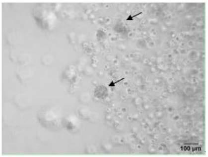
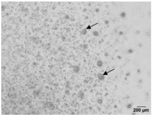
Moreover, there have been no reports on the use of 3D culture technology to explore the lactation function of porcine mammary epithelial cells at home and abroad.
Method used
Image
Click on the blue label to locate the original text in one second.
Reading with bidirectional positioning of images and text.
Examples
Embodiment 1
[0028] The present embodiment provides a method for culturing porcine mammary epithelial cells in 3D, comprising the following steps:
[0029] (1) Preheat the 24-well plate at 37°C;
[0030] (2) Digest pig mammary gland epithelial cells before generation 10 into single cells with trypsin and centrifuge at 1000rpm for 4min, discard the supernatant, and keep the precipitate;
[0031] (3) Resuspend the precipitate in a mixed solution prepared at 5°C with Matrigel and Advanced DMEM / F12 complete medium at a volume ratio of 1:1, and adjust the concentration of porcine mammary gland epithelial cells to 4×10 4 cell / ml;
[0032] (4) Drop the resuspension solution into the 24-well plate according to the amount of 30-40ul per drop, drop one drop into each well, incubate the 24-well plate at 37°C for 40min to form a gel;
[0033] (5) Add 550ul Advanced DMEM / F12 complete medium to the 24-well plate and continue culturing for 2 days, and replace the Advanced DMEM / F12 complete medium every...
Embodiment 2
[0037] The present embodiment provides a method for culturing porcine mammary epithelial cells in 3D, comprising the following steps:
[0038] (1) Preheat the 6-well plate at 37°C;
[0039] (2) Digest pig mammary gland epithelial cells before generation 10 into single cells with trypsin and centrifuge at 1200rpm for 4min, discard the supernatant, and keep the precipitate;
[0040] (3) Resuspend the precipitate in the mixed solution prepared by Matrigel and Advanced DMEM / F12 complete medium at a volume ratio of 1:1 at 6°C, and adjust the concentration of porcine mammary gland epithelial cells to 1×10 4 cell / ml;
[0041](4) Drop the resuspension solution into a 6-well plate according to the amount of 30-40ul per drop, drop 6 drops into each well, incubate the 6-well plate at 37°C for 40 minutes to form a gel;
[0042] (5) Add 2 mL of Advanced DMEM / F12 complete medium to the 6-well plate to continue culturing for 3 days, and replace the Advanced DMEM / F12 complete medium every o...
PUM
Abstract
The invention provides a method for culturing porcine mammary epithelial cells by 3D. The method comprises the following steps: (1) preheating a pore plate at 37 DEG C; (2) digesting porcine mammary epithelial cells of generations earlier than the 10th generation into single cells by trypsin, performing centrifugation, removing supernatant, and retaining precipitate; (3) resuspending the precipitate with a mixed solution prepared from Matrigel and an Advanced DMEM / F12 complete medium at 0-10 DEG C in a volume ratio of 1:1, and adjusting the concentration of the porcine mammary epithelial cellsto 1*10<4>-5*10<4> cells / ml; (4) adding dropwise the resuspension solution according to 30-40 mul per drop to the pore plate, and inverting the pore plate for incubation at 37 DEG C for 30-40 min toform gel; (5) adding the Advanced DMEM / F12 complete medium to the pore plate for continuous culture for 2-3 days, and replacing the Advanced DMEM / F12 complete medium once every other day.
Description
technical field [0001] The invention relates to the technical field of cell culture, in particular to a method for culturing porcine mammary epithelial cells in 3D. Background technique [0002] 3D culture technology can better simulate the microenvironment in vivo, better reflect the polarized morphology of cells, and the interaction between cells and between cells and matrix. 3D culture is to cultivate cells in a certain extracellular matrix (ECM). The ECM protein acts as a growth scaffold, enabling cells to differentiate to produce a certain three-dimensional tissue-specific structure. The cell growth environment created simulates the in vivo environment to the greatest extent. . There are five main methods of 3D culture technology: (a) hanging drop method; (b) rotating system; (c) stirring benchmark method; (d) matrix and scaffold; (e) microfluidic system [1] . Researchers at home and abroad mainly use Matrigel as a scaffold for 3D culture. Matrigel is a basement mem...
Claims
Application Information
Patent Timeline
Application Date:The date an application was filed.
Publication Date:The date a patent or application was officially published.
First Publication Date:The earliest publication date of a patent with the same application number.
Issue Date:Publication date of the patent grant document.
PCT Entry Date:The Entry date of PCT National Phase.
Estimated Expiry Date:The statutory expiry date of a patent right according to the Patent Law, and it is the longest term of protection that the patent right can achieve without the termination of the patent right due to other reasons(Term extension factor has been taken into account ).
Invalid Date:Actual expiry date is based on effective date or publication date of legal transaction data of invalid patent.

