Medical image processing method, device, system and storage medium

A medical image and processing method technology, applied in the field of neural network, can solve problems such as poor auxiliary effect, good diagnosis basis for Parkinson's disease diagnosis, etc., and achieve the effect of improving accuracy.

Inactive Publication Date: 2019-01-18
SHANGHAI UNITED IMAGING INTELLIGENT MEDICAL TECH CO LTD
View PDF9 Cites 34 Cited by
  • Summary
  • Abstract
  • Description
  • Claims
  • Application Information

AI Technical Summary

Problems solved by technology

[0005] Based on this, it is necessary to provide a medical image processing method, device, system and storage medium for the problem that the ...

Method used


Image

  • Medical image processing method, device, system and storage medium
  • Medical image processing method, device, system and storage medium
  • Medical image processing method, device, system and storage medium

Examples


Embodiment Construction

[0057] In order to make the purpose, technical solutions and advantages of the present invention clearer, the technical solutions in the embodiments of the present invention will be further described in detail through the following embodiments in conjunction with the accompanying drawings. It should be understood that the specific embodiments described here are only used to explain the present invention, not to limit the present invention.

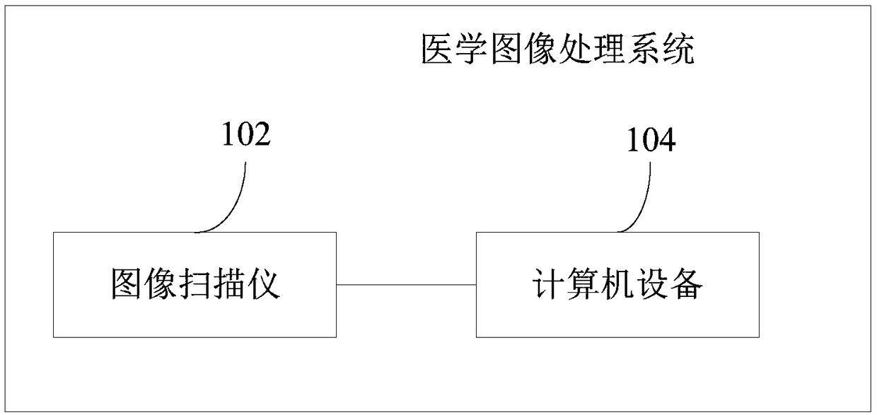
[0058] The medical image processing method provided by the present invention can be applied to such as figure 1 The medical image processing system shown. The system includes an image scanner 102 and a computer device 104 . The image scanner 102 can be a magnetic resonance scanner, or a CT scanner, etc. The image scanner 102 is used to acquire detection images, and the computer device 104 can be a terminal, such as a personal computer, a notebook, and a tablet computer. The computer device 104 includes a processor, memory, network interf...


PUM


Abstract

The invention relates to a medical image processing method, a device, a system and a storage medium. The method includes acquiring a detection image including an image of a substantia nigra region and/or a red nucleus region; Obtaining a segmentation neural network model, and performing segmentation operation on the detection image by adopting the segmentation neural network model to obtain a target mask image; obtaining a detection neural network model, and obtaiing the detection result by detecting the target mask image using the detection neural network model. This method can make more accurate detection results of detection images according to the trained detection neural network model, and further improve the accuracy of detection results.

Description

technical field [0001] The present invention relates to the technical field of neural networks, in particular to a medical image processing method, device, system and storage medium. Background technique [0002] Parkinson's disease (Parkinson's disease, PD) is a common neurodegenerative disease. For patients diagnosed with Parkinson's disease, the life expectancy is about 7 to 14 years. Therefore, the diagnosis of Parkinson's disease is very critical for later treatment. The clinical diagnosis of Parkinson's disease mainly relies on the patient's medical history, clinical symptoms, and drug reactions, and brain imaging is used to assist in the diagnosis of Parkinson's disease. [0003] In the traditional technology, the MRI T1 image of the brain of the diagnosed object is mainly obtained by using Magnetic Resonance Imaging (MRI) T1-weighted imaging technology. The MRI T1 image mainly reflects the brain structure. By comparing the brain structure with the brain structure of ...

Claims


Application Information

Patent Timeline
IPC IPC(8): G06T7/00G06T7/11G06N3/08
CPCG06N3/08G06T7/0012G06T2207/10088G06T7/11
Inventor 石峰詹翊强
Owner SHANGHAI UNITED IMAGING INTELLIGENT MEDICAL TECH CO LTD
Why Eureka
Try Eureka