
A method and apparatus for image region segmentation of ischemic stroke

An ischemic stroke and image area technology, applied in the field of image processing, can solve problems such as process dispersion, segmentation results are easily affected, and interference, and achieve the effects of avoiding errors, improving segmentation accuracy, and saving labor costs

Active Publication Date: 2019-01-18
BEIJING SENSETIME TECH DEV CO LTD
View PDF8 Cites 9 Cited by
  • Summary
  • Abstract
  • Description
  • Claims
  • Application Information

AI Technical Summary

Problems solved by technology

The whole process is relatively scattered, and there are too many interferences of human factors, the final segmentation results are more easily affected

Method used


Image

  • A method and apparatus for image region segmentation of ischemic stroke
  • A method and apparatus for image region segmentation of ischemic stroke
  • A method and apparatus for image region segmentation of ischemic stroke

Examples


Embodiment Construction

[0026] Embodiments of the present application are described below with reference to the drawings in the embodiments of the present application.

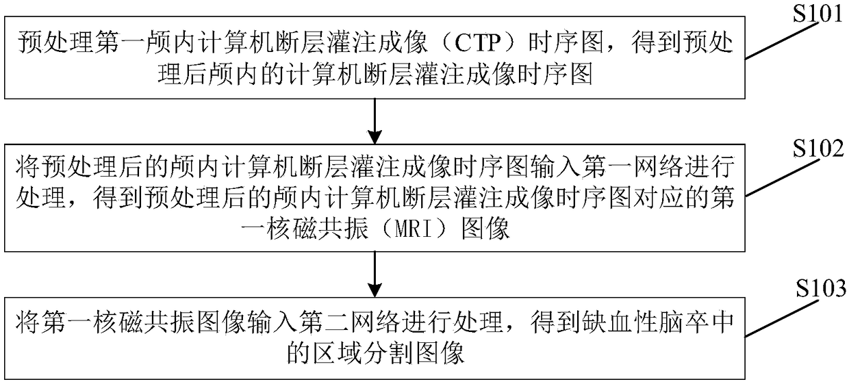
[0027] see figure 1 , figure 1 is a schematic flow chart of a method for segmenting an ischemic stroke image region provided in an embodiment of the present application.

[0028] S101. Preprocessing the first intracranial computed tomography perfusion imaging (CTP) timing diagram to obtain a preprocessing intracranial computed tomography perfusion imaging timing diagram.

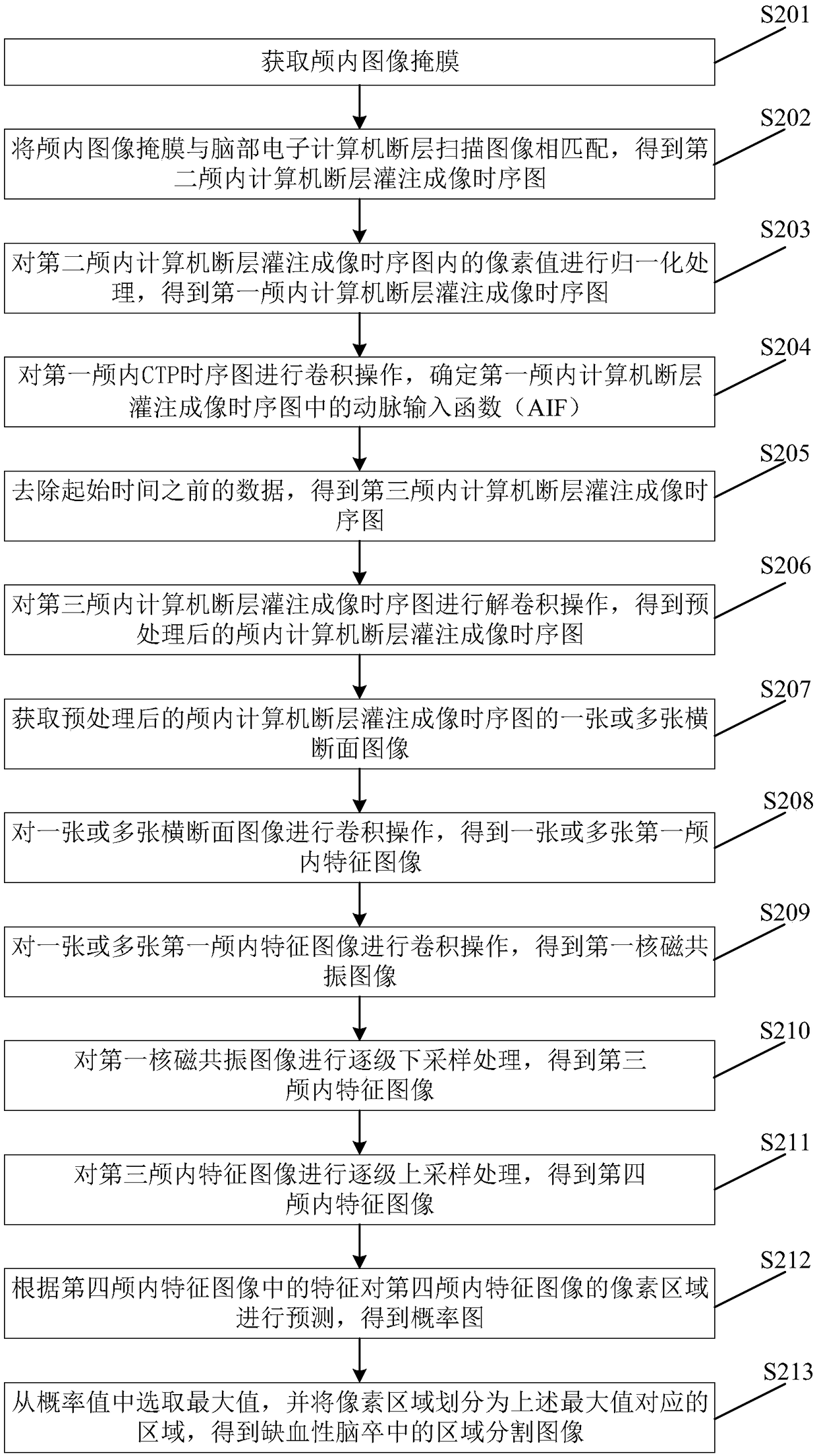
[0029] According to the computerized tomography image of the brain obtained by CT scanning, the CTP timing diagram can be obtained, and the subsequent distinction between the cerebral infarction area and the penumbra area is based on the CTP timing diagram, but the CTP timing diagram contains a large number of Invalid data, the existence of these invalid data will reduce the final segmentation accuracy and directly affect the doctor's diagnosis of the patient. T...


PUM


Abstract

A method and apparatus for image region segmentation of ischemic stroke are disclosed. The method comprises the following steps: preprocessing the CTP sequence diagram of the first intracranial computed tomography perfusion imaging to obtain the CTP sequence diagram of the intracranial after preprocessing; Inputting the preprocessed intracranial CTP timing diagram into a first network for processing to obtain a first nuclear magnetic resonance MRI image corresponding to the preprocessed intracranial CTP timing diagram; inputting the first MRI image into a second network for processing to obtain a region segmentation image of ischemic stroke. A corresponding device is also disclosed. Through a series of convolution operations on the CTP timing diagram, the CTP timing diagram is automatically and efficiently divided into cerebral infarction region, penumbra region and background region. The whole process does not require manual participation, which can effectively avoid the error causedby manual participation, save human cost, and can efficiently and fast complete the CTP timing diagram distinction.

Description

technical field [0001] The present application relates to the technical field of image processing, in particular to a method and device for ischemic stroke image region segmentation. Background technique [0002] Stroke, also known as "stroke", is an acute cerebrovascular disease with a very high fatal risk. Includes ischemic and hemorrhagic stroke. The incidence of ischemic stroke is higher than that of hemorrhagic stroke, accounting for 60%-70% of the total stroke. After the onset of ischemic stroke, an ischemic penumbra will be formed around the infarction after the occurrence of cerebral infarction. There are still a large number of dormant and semi-dormant brain cells in the penumbra. Therefore, how to save the penumbra becomes the key to clinical treatment. CTP (Computed Tomography Perfusion) has begun to be used as an early diagnosis method for ischemic stroke due to some excellent characteristics (such as: fast imaging speed, easy acquisition, etc.), and it can co...

Claims


Application Information

Patent Timeline
Patent Type & Authority Applications(China)
IPC IPC(8): G06T7/11G06N3/04
CPCG06T7/11G06T2207/30016G06T2207/10081G06N3/045
Inventor 刘蓬博宋涛
Owner BEIJING SENSETIME TECH DEV CO LTD
Learn More