

Oral cavity defect repair membrane and preparation method thereof
A technology of mucosal layer and submucosa layer, applied in medical science, tissue regeneration, prosthesis, etc., can solve problems such as unreasonable preparation methods, unsuitable oral defect repair, unsuitable animal-derived materials, etc., and achieve a good tissue microporous structure and mechanical properties, avoiding the loss of cell growth factors, avoiding the effects of cytotoxicity
- Summary
- Abstract
- Description
- Claims
- Application Information
AI Technical Summary
Benefits of technology
Problems solved by technology
Method used
Image


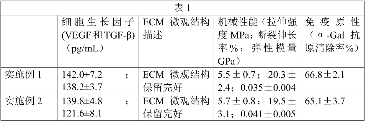
Examples
Embodiment 1
[0027] This embodiment provides a porcine small intestinal submucosa tissue acellular matrix material and a preparation method thereof. The preparation method includes:
[0028] Step 1. Take the porcine small intestine jejunum section, clean it, cut it radially, and wash it repeatedly with PBS solution (phosphate buffered saline solution) to remove pollutants.
[0029] Step 2. Physically remove the mucous membrane, using a wooden scraper, carefully and evenly scrape off the mucous membrane layer of the inner layer of the SIS membrane.
[0030] Step 3. Physically remove the sarcolemma and serosa. The wooden scraper and scalpel are used together to remove the sarcolemma and serosa on the outer layer of the SIS membrane.
[0031] Step 4, configuring 0.1% (mass fraction) peracetic acid solution, and then soaking the SIS membrane with the mucous membrane layer, sarcolemma layer and serosa layer removed in 0.1% peracetic acid solution for 7 hours.
[0032] Step 5, taking out the S...
Embodiment 2
[0038] This embodiment provides a porcine small intestinal submucosa tissue acellular matrix material and a preparation method thereof. The preparation method includes:
[0039] Step 1. The jejunum section of the porcine small intestine was taken, cleaned and cut radially, and washed repeatedly with PBS solution to remove pollutants.
[0040] Step 2. Physically remove the mucous membrane, using a wooden scraper, carefully and evenly scrape off the mucous membrane layer of the inner layer of the SIS membrane.
[0041] Step 3. Physically remove the sarcolemma and serosa. The wooden scraper and scalpel are used together to remove the sarcolemma and serosa on the outer layer of the SIS membrane.
[0042] Step 4, configuration 1% (mass fraction) sodium hydroxide solution and the TritonX-100 solution of 0.5% (mass fraction), will remove the SIS membrane of mucous membrane layer, sarcolemma and serosal layer and soak in 0.1% (mass fraction) Sodium hydroxide solution for 7 hours.
...
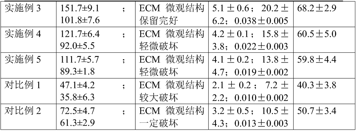
Embodiment 3
[0050] This embodiment provides a porcine small intestinal submucosa tissue acellular matrix material and a preparation method thereof. The preparation method includes:
[0051] Step 1. Wash and cut radially, and rinse repeatedly with PBS solution to remove contaminants.
[0052] Step 2. Physically remove the mucous membrane, using a wooden scraper, carefully and evenly scrape off the mucous membrane layer of the inner layer of the SIS membrane.
[0053] Step 3. Physically remove the sarcolemma and serosa. The wooden scraper and scalpel are used together to remove the sarcolemma and serosa on the outer layer of the SIS membrane.
[0054] Step 4, preparing 1% sodium hydroxide solution, and then soaking the SIS membrane with the mucous membrane layer, sarcolemma layer and serosa layer removed in 0.1% sodium hydroxide solution for 7 hours.
[0055] Step 5, taking out the SIS membrane treated with sodium hydroxide solution, and washing it repeatedly with PBS solution for 3 times...
PUM
 Abstract
Description
Claims
Application Information