Medical device for medical image analysis

A technology of medical equipment and medical imaging, which is applied in the field of medical equipment for medical image analysis, can solve the problem of inconvenient adjustment of background light, and achieve an effect that is conducive to observation and analysis

Pending Publication Date: 2019-01-29
翟树校
View PDF0 Cites 4 Cited by
  • Summary
  • Abstract
  • Description
  • Claims
  • Application Information

AI Technical Summary

Problems solved by technology

[0004] The purpose of the present invention is to provide a medical device for medical image analysis. The torch installed on the back of the light box through a metal hose can irradiate the loc

Method used


Image

  • Medical device for medical image analysis
  • Medical device for medical image analysis
  • Medical device for medical image analysis

Examples


Embodiment Construction

[0022] The following will clearly and completely describe the technical solutions in the embodiments of the present invention with reference to the accompanying drawings in the embodiments of the present invention. Obviously, the described embodiments are only some, not all, embodiments of the present invention. Based on the embodiments of the present invention, all other embodiments obtained by persons of ordinary skill in the art without making creative efforts belong to the protection scope of the present invention.

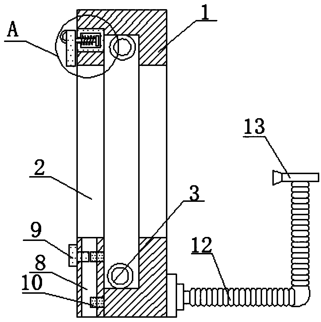
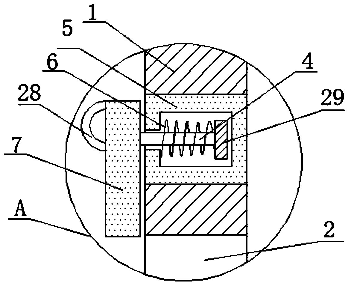
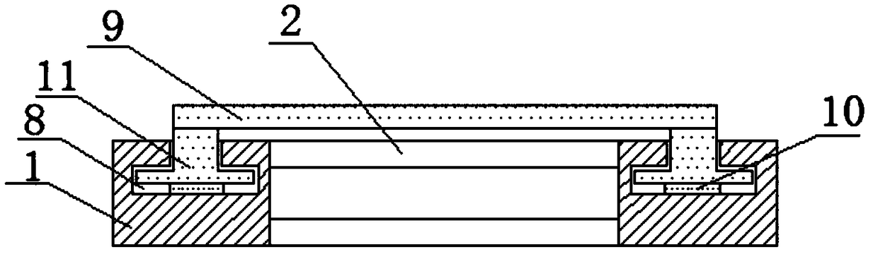
[0023] see Figure 1~6 , the present invention provides a technical solution: a medical device for medical image analysis, including a light box 1, a transparent sheet 2 is installed on both sides of the light box 1, and the upper and lower ends of the transparent sheet 2 are installed with The lighting tube 3, the upper end of one side of the light box 1 is provided with a convex groove 5, the interior of the convex groove 5 is movably connected to a limiting...


PUM


Abstract

The invention discloses a medical device for medical image analysis. The medical device includes a light box; transparent sheets are arranged on two sides of the light box; illumination tubes are arranged on inner upper and lower ends of the transparent sheets; a concave groove is formed in an upper end of one side of the light box; a limiting plate is movably connected in the concave groove; oneside of the limiting plate is fixedly connected to a linkage rod; one end of the linkage rod is fixedly connected to a first clamping plate; T-shaped sliding grooves are formed in the lower end of oneside of the light box; T-shaped sliding blocks are movably connected in the T-shaped sliding grooves; the tops of the T-shaped sliding blocks are fixedly connected with a second clamping plate; magnets are arranged on the bottoms of the T-shaped sliding grooves; and a metal hose is arranged on the other side of the light box, and a flashlight is arranged on one end of the metal hose. The flashlight arranged on the back of the light box through the metal hose can locally irradiate the CT piece, and facilitates observation and analysis.

Description

technical field [0001] The invention relates to the technical field of medical machinery, in particular to a medical device for medical image analysis. Background technique [0002] At present, for the needs of clinical diagnosis, patients will collect and check the images of various medical imaging equipment (such as CT, MRI, ultrasound, etc.) on the tissues and organs of the same body part in the medical imaging examination, and judge the completion effect of interventional ablation surgery , it is necessary to analyze whether the ablation area is complete. Therefore, it is necessary to collect images of a certain range around the patient's treatment area before and after surgery, and then undergo appropriate image segmentation and registration to match the image content and position, so as to view the treatment effect. [0003] Now medical staff use traditional plastic CT films, which are relatively large and easy to carry and store, and are easy to be damaged due to acci...

Claims


Application Information

Patent Timeline
IPC IPC(8): G02B27/02
CPCG02B27/024G02B27/028
Inventor 翟树校
Owner 翟树校
Why Eureka
Try Eureka