A brain anatomy teaching system based on magnetic resonance imaging

A teaching system, magnetic resonance technology, applied in the field of brain anatomy teaching system, can solve the problem that the digital three-dimensional model structure cannot be confirmed by the correlation of the two-dimensional image structure, cannot meet the requirements of modern medical teaching, and the separation of anatomy and imaging teaching increases learning Difficulty and other issues

Pending Publication Date: 2019-02-05
迈格生命科技(深圳)有限公司
View PDF4 Cites 9 Cited by
  • Summary
  • Abstract
  • Description
  • Claims
  • Application Information

AI Technical Summary

Problems solved by technology

[0004] Using cadaver specimens for anatomy teaching is the most intuitive, but due to limited specimen resources, it cannot meet the current teaching quantity needs
The use of brain atlas images for teaching is the most cost-effective and the most widely used method. However, since the current human brain anatomy and imaging education are separated, students need to use their own imagination to create two-dimensional images and three-dimensional brain images during the learning process. The correlation between structures, the identification of some fine brain regions in the image itself is very difficult, and the separation of anatomy and imaging teaching makes learning

Method used


Image

  • A brain anatomy teaching system based on magnetic resonance imaging
  • A brain anatomy teaching system based on magnetic resonance imaging
  • A brain anatomy teaching system based on magnetic resonance imaging

Examples


no. 1 example

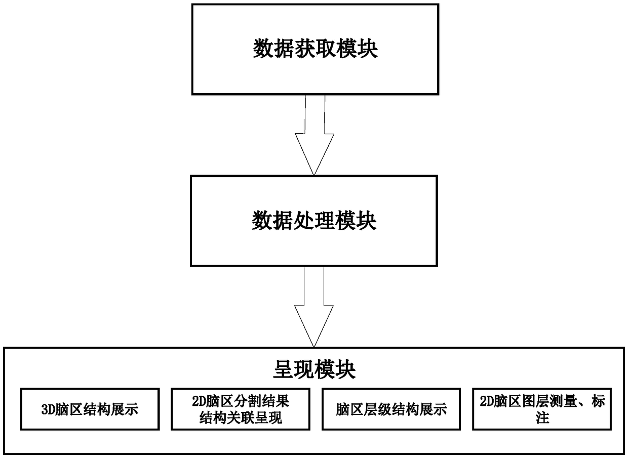
[0030] This example is randomly collected brain magnetic resonance imaging data of any individual. The original brain magnetic resonance image data provides three sets of standard DICOM grayscale image files of cross section, sagittal plane and coronal plane.

[0031] After obtaining the data file, we use the brain atlas method to segment the brain region. The method is to use registration to map the image to be segmented to the template that has been segmented, and then transform the segmentation result to the original image through the mathematical inverse transformation operator. space, so as to obtain the segmentation result of the original input image. Using these segmentation results for 3D reconstruction, the 3D model data of a single brain region can be obtained. Using a fixed calculation method, each brain region can be independently analyzed and related quantitative data can be obtained.

[0032]The purpose of the large deformation differential homeomorphism (LDDMM)...


PUM


Abstract

The invention provides a brain anatomy teaching system based on magnetic resonance image, By importing the MRI data of real cases which had been segmented and reconstructed automatically in advance, In order to realize one-key interaction between two-dimensional images and three-dimensional brain anatomy, two-dimensional segmentation structure display and digital three-dimensional model demonstration of any single brain region and multi-brain regions are provided, which provides a scheme for the teaching of brain anatomy based on real cases.

Description

technical field [0001] The invention belongs to the field of medical image processing technology and medical teaching, in particular to a brain anatomy teaching system based on magnetic resonance images. Background technique [0002] With the in-depth application of computer technologies such as medical imaging data collection and digital image processing in the medical field, digital teaching has gained a higher and higher status in medical teaching, and has achieved rapid development in recent years. As an important part of the medical field, medical teaching is gradually shifting towards digitization and virtualization. In medical teaching and scientific research, human anatomy is an important basic course, and it is the only way to learn and understand human body structure and medical research. [0003] The existing forms of human brain anatomy teaching include the use of cadaver specimens for anatomy practice, the use of atlas images for brain tissue demonstration, and...

Claims


Application Information

Patent Timeline
IPC IPC(8): G06T3/00G06T7/11
CPCG06T3/005G06T7/11G06T2207/10088G06T2207/10012G06T2207/30016
Inventor 马婷吕海岩徐鹏
Owner 迈格生命科技(深圳)有限公司
Why Eureka
Try Eureka