
A method for detecting the rigid exudate lesion in a fundus image based on a convolution neural network

A convolutional neural network and fundus image technology, applied in image enhancement, image analysis, image data processing, etc., can solve the problems of low screening efficiency and low algorithm accuracy, achieve good detection results, simplify the image processing process, The effect of enhancing contrast

Inactive Publication Date: 2019-03-08
TIANJIN POLYTECHNIC UNIV
View PDF4 Cites 11 Cited by
  • Summary
  • Abstract
  • Description
  • Claims
  • Application Information

AI Technical Summary

Problems solved by technology

In view of the shortcomings of low efficiency of fundus lesions screening in hospitals and low accuracy of traditional algorithms, applying deep learning methods to the detection of hard exudates in fundus images is of great significance for improving the accuracy of detection of hard exudates in fundus images

Method used


Image

  • A method for detecting the rigid exudate lesion in a fundus image based on a convolution neural network
  • A method for detecting the rigid exudate lesion in a fundus image based on a convolution neural network
  • A method for detecting the rigid exudate lesion in a fundus image based on a convolution neural network

Examples


Embodiment Construction

[0027] The present invention will be further described in detail below in combination with specific embodiments.

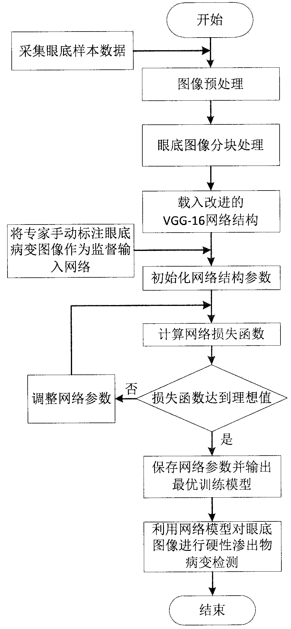
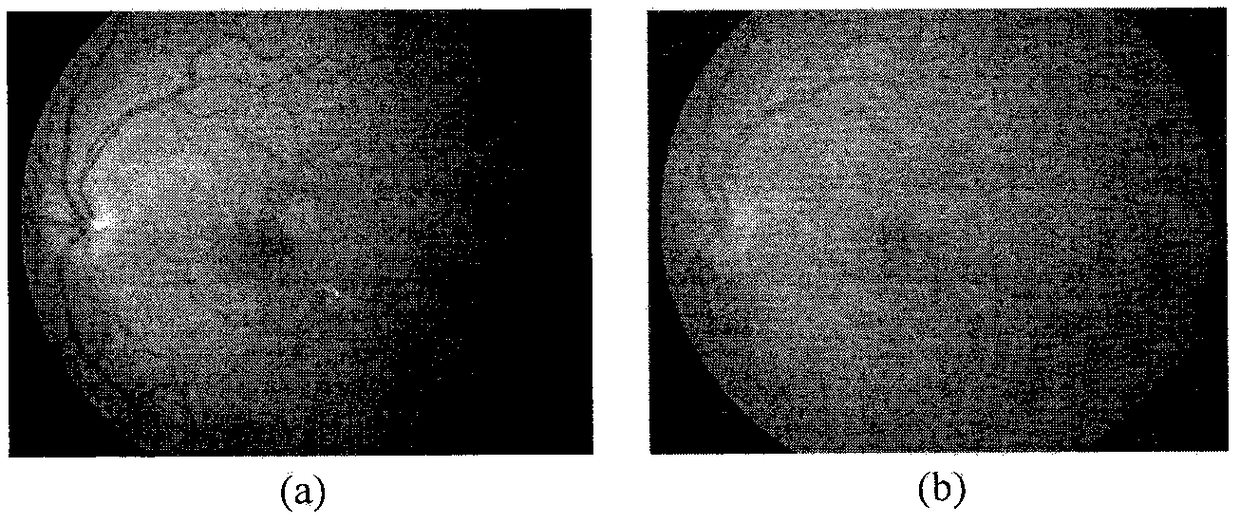
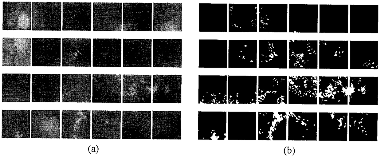
[0028] The overall framework schematic diagram of the present invention is as figure 1 As shown, firstly, the images collected by DRIVE, MESSIDOR database and fundus camera are preliminarily sorted out, experts manually label and grade the lesions, and the contrast of the images is enhanced through Gamma correction to make the images more suitable for network training; the preprocessed images are normalized and cropped To adapt to the scale of network training data and enhance accuracy; improve the VGG-16 network structure by introducing channel weighting structure, multi-scale feature structure and optimized residual module, in which the original image in the training image is a normalized fundus image, The fundus images manually marked by experts are used as training supervision images, which are input into the optimized VGG-16 network, and the network parameter...


PUM


Abstract

The invention provides a method for detecting the rigid exudate lesion in a fundus image based on a convolution neural network. The method comprises the following steps of 1) preprocessing the fundusimage and enhancing image contrast through Gamma correction algorithm; 2) carrying out block processing on that image to expand the data; 3) using the lesion results manually annotated by experts in DRIVE, MESSIDOR database as the monitoring data, using the adjusted VGG-16 network to train the block image and introducing the channel weighting module to establish the dependency of the feature channel explicitly by learning; 4) using the trained network model to detect the hard exudate lesion in the fundus image. Compared with the traditional scheme, the method of the invention avoids the complicated image processing process, is not affected by the fundus optic disc and cotton floc spots on the hard exudate lesion, realizes the high-precision detection of the hard exudate lesion, and can bewidely used in the field of automatic screening of the fundus rigid exudate.

Description

technical field [0001] The invention relates to a method for detecting hard exudates in fundus images based on convolutional neural networks, which is better than traditional algorithms in terms of sensitivity, specificity and accuracy, and has better detection performance for hard exudates in fundus images. processing, medical lesion detection, and deep learning. Background technique [0002] Diabetic retinopathy (Diabetic retinopathy, DR) is the most common eye complication of diabetes, and it is the main cause of new blindness in adults aged 20-65. Most of the people who felt uncomfortable and went to see a doctor were already visually impaired. Therefore, early detection of retinopathy is very important to reduce the chance of blindness in the eyes of patients. However, at present, there are few medical ophthalmologists, the inspection efficiency is low, and comprehensive screening cannot be performed. The computer-aided screening system is a favorable tool to improve ...

Claims


Application Information

Patent Timeline
IPC IPC(8): G06T7/00G06T5/00
CPCG06T7/0012G06T2207/20084G06T2207/20081G06T2207/30041G06T5/92
Inventor 张芳肖志涛徐旭耿磊吴骏王雯刘彦北
Owner TIANJIN POLYTECHNIC UNIV
Learn More