Enhanced gene delivery methods

A technology of gene delivery and target cells, applied in other methods of inserting foreign genetic materials, biochemical equipment and methods, pharmaceutical formulations, etc.

Pending Publication Date: 2019-03-15
ANGIOCRINE BIOSCI
View PDF7 Cites 1 Cited by
  • Summary
  • Abstract
  • Description
  • Claims
  • Application Information

AI Technical Summary

Problems solved by technology

Unfortunately, attempts at gene correction in these and other cells have so far proven largely ineffective

Method used


Image

  • Enhanced gene delivery methods
  • Enhanced gene delivery methods
  • Enhanced gene delivery methods

Examples


Embodiment 1

[0084] Effect of E4ORF1+ co-culture on HSPC expansion and virus transduction

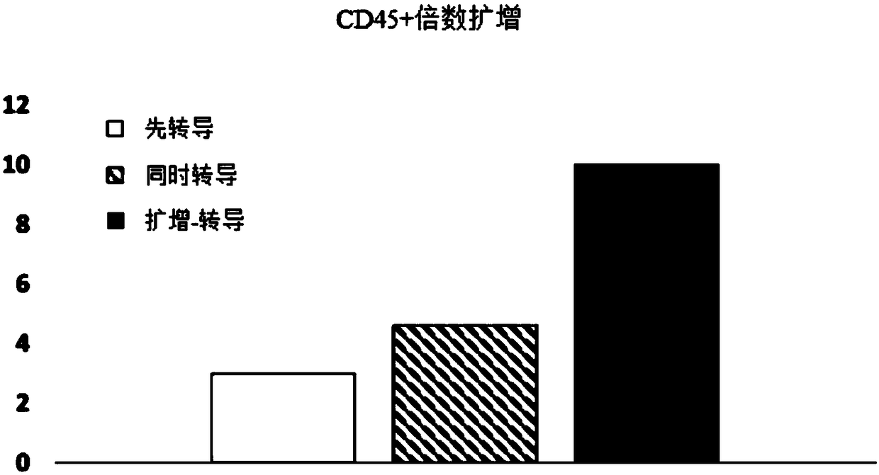
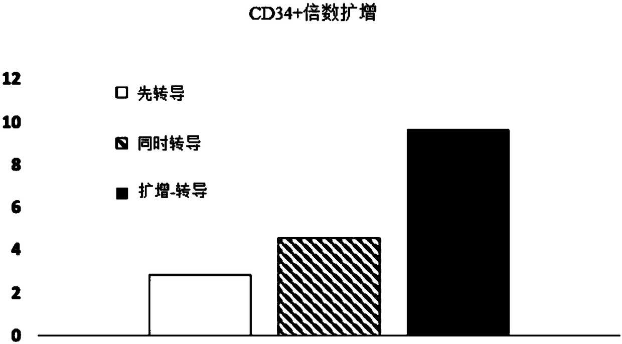
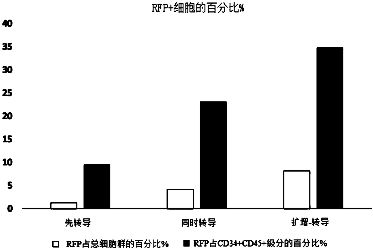
[0085] Experiments were performed to assess the effect of E4ORF1+ endothelial cells on the ability to expand and transduce CD34+ / CD45+ HSPCs. CD34+ cells (Lonza) were transduced with red fluorescent protein (RFP) and expanded on E4ORF1+ umbilical vein endothelial cell (UVEC) cultures in 6-well plates according to three different protocols as follows:

[0086] In protocol 1, transduction was initiated 24 hours before initiation of HSPC-E4ORF1+UVEC co-culture ("transduction first").

[0087] In protocol 2, transduction was performed while co-cultivating with HSPC-E4ORF1+UVEC (simultaneous transduction).

[0088] In protocol 3, HSPCs were co-cultured with E4ORF1+UVECs for 48 hours, after which the floating HSPCs were transferred to empty wells for transduction (24 hours), after transduction, HSPCs were then co-cultured with E4ORF1+UVECs ("amplification-transduction").

[0089] In each of the above...

Embodiment 2

[0092] Suspension Fraction & Cytokines Effect on Viral Transduction Efficiency

[0093] Experiments were performed to determine the effect of removal of the "floating fraction" of CD34+ hematopoietic cells on transduction efficiency, and to determine whether cytokine concentration had an effect on transduction efficiency.

[0094] CD34+ cells (Lonza) were transduced with blue fluorescent protein (BFP) and expanded on E4ORF1+UVEC cultures in 6-well plates according to four different protocols as follows:

[0095] In protocols 1 and 3, HSPCs were expanded on E4ORF1+UVEC cultures for 96 hours and subsequently transduced by incubating the HSPCs with a lentivirus engineered to express BFP for 24 hours, while the HSPCs were still on the E4ORF1+EC feeder layer superior. Then, HSPCs were expanded for two more days in E4ORF1+EC cultures. The results obtained using these protocols are referred to in the figure as "co-transduction" results.

[0096] In protocols 2 and 4, HSPCs were ...

Embodiment 3

[0102] Rescue of transfected cells by subsequent EC co-culture

[0103] Transfected or transduced cells, eg, cells transfected by electroporation, can be cultured with ECs after transfection or transduction. This subsequent co-cultivation can "rescue" cells that were destroyed during transfection or transduction - reducing death and loss of transfected or transduced cells. This co-cultivation should start "immediately" (as defined in this patent specification) after the start of the transfection or transduction step, and should continue for a sufficient time to allow recovery of the target cells. Without wishing to be bound by theory, it is believed that such "rescue" may be the result of cell-cell contact between the two cell populations (i.e. transduced / transfected cells and EC), and / or may be the result of transducing Exposure of transfected cells results in soluble factors secreted by ECs, such as angiogenic factors and cytokines. Co-cultivation of the two cell populat...


PUM


Abstract

The invention provides improved methods for gene delivery to, or genetic modification of target cells, wherein the gene delivery or other genetic modification of the target cells is performed in the presence of endothelial cells, or after co-culture of the target cells with endothelial cells, or wherein co-culture of the target cells with endothelial cells is employed immediately alter gene delivery in order to "rescue" cells that may have been damaged during the gene delivery process. In some embodiments gene delivery is performed by transfection. In some embodiments gene delivery is performed by transduction, in some embodiments the endothelial cells are organ-specific endothelial cells. In some embodiments the endothelial cells are E4ORF1-expressing endothelial cells (E4ORF1+ECs). In some embodiments the target cells are stem cells, such as hematopoietic stem cells.

Description

[0001] Cross References to Related Applications [0002] This application claims priority to U.S. Provisional Patent Application No. 62 / 323,476, filed April 15, 2016, and U.S. Provisional Patent Application No. 62 / 403,110, filed October 1, 2016, the contents of which are hereby incorporated by reference The method is incorporated into this article as a whole. [0003] incorporated by reference [0004] For use only in those jurisdictions that permit incorporation by reference, all references cited in this application (including but not limited to publications, patent applications, patents, and other references) are hereby incorporated by reference in their entirety in their entirety This article. In addition, any manufacturer's instructions or catalogs for any products cited or mentioned herein are hereby incorporated by reference. Many of the teachings provided in US Patent No. 8,465,732 can be used in conjunction with, or can be adapted to, the present invention. According...

Claims


Application Information

Patent Timeline
Patent Type & Authority Applications(China)
IPC IPC(8): A61K35/545A61K35/28A61K35/44C12N5/0789C12N5/10
CPCA61K35/28A61K35/44A61K35/545C12N15/86C12N2740/16043C12N15/87
Inventor 保罗·威廉·芬尼根克劳德·杰弗里·戴维斯迈克尔·丹尼尔·金斯伯格丹尼尔·约瑟夫·诺兰
Owner ANGIOCRINE BIOSCI
Why Eureka
Try Eureka